乳糖/D-半乳糖[快速]检测试剂盒 Lactose/Sucrose/D-Glucose Assay Kit 货号:K-LACGAR Megazyme中文站

乳糖/D-半乳糖[快速]检测试剂盒

英文名:Lactose/Sucrose/D-Glucose Assay Kit

货号:K-LACGAR

规格:115 assays per kit

市场价: 5700

乳糖/D-半乳糖[快速]检测试剂盒

Megazyme低聚半乳糖和半乳糖检测试剂盒采用专利技术,在试剂盒中加入半乳糖变旋酶,可快速催化限速变旋步骤,室温下5分钟即可获得检测结果。试剂盒规格:115次方法:分光光度计,340nm 反应时间: 5分钟检测限: 2.96mg/L 样品类型:牛奶、乳制品(如奶油、奶粉、乳清粉、奶酪、炼乳和酸奶)、含乳食品(如保健食品、焙烤食品、婴幼儿食品、巧克力、糖果和冰淇淋)、食品添加剂、饲料、化妆品、医药及其他物料。方法认证: 通过AOAC、NBN、DIN、GOST以及德国、荷兰、瑞士和奥地利的认证.

分析物意义:常见加工食品组分,在某些情况下,精确的数值很重要,如 “无乳糖”产品 

Megazyme检测试剂盒优点:K-LACGAR试剂盒反应快(室温,5min)、试剂稳定

The Lactose/Galactose (Rapid) test kit is used for the rapid test of lactose, D-galactose and L-arabinose in food and plant products. Galactose dehydrogenase can be used the measurement and analysis of both D-galactose and L-arabinose. Suitable for the analysis of lactose in “low-lactose” or “lactose-free” samples which contain high levels of monosaccharides.

UV-method for the determination of Lactose and D-Galactose in
foodstuffs, beverages and other materials

Principle:
(β-galactosidase)
(1) Lactose + H2O → β-D-galactose + D-glucose

(galactose mutarotase)
(2) α-D-Galactose ↔ β-D-galactose

(β-galactose dehydrogenase)
(3) β-D-Galactose + NAD+ → D-galactonic acid + NADH + H+
 

Kit size: 115 assays
Method: Spectrophotometric at 340 nm
Reaction time: ~ 15 min
Detection limit: 2.96 mg/L (lactose)
Application examples:
Milk, dairy products (e.g. cream, milk / whey powder, cheese,
condensed milk and yogurt), foods containing milk (e.g. dietetic foods,
bakery products, baby food, chocolate, sweets and ice-cream), food
additives, feed, cosmetics, pharmaceuticals and other materials
(e.g. biological cultures, samples, etc.)
Method recognition:
Methods based on this principle have been accepted by AOAC, NBN,
DIN, GOST and IDF

Advantages

  • Very rapid reaction due to inclusion of galactose mutarotase (patented technology PCT / IE2004 / 00170)
  • Very competitive price (cost per test)
  • All reagents stable for > 2 years after preparation
  • Mega-Calc™ software tool is available from our website for hassle-free raw data processing
  • Standard included

Q1. Should the pH of the sample be adjusted even for samples in acidic media?

The pH of the assay solution after the sample is added should be the same as that of the assay buffer that is supplied with the kit.
Low sample volumes (e.g. 0.1 mL) are not likely to affect the pH of the assay solution and therefore may not require pH adjustment.
Samples above 0.1 mL are more likely to affect the pH of the assay solution and therefore the pH of these samples should be adjusted as described in the data booklet, prior to addition to the assay.

Q2. Why is the borohydride reduction step required in procedure B?

Procedure B is required  for  “low-lactose” or “lactose-free” samples containing high levels of monosaccharides.  Generally, these types of samples contain high levels of  “free” galactose which causes a high background and reduces the dynamic range available to measure the galactose that is released from lactose in the test.  To avoid this the borohydride step is used to reduce the free galactose.

Q3. Sometimes a negative absorbance change is obtained for the blank samples, is this normal? Should the real value (negative absorbance change) or “0” be used in the calculation of results?

Sometimes the addition of the last assay component can cause a small negative absorbance change in the blank samples due to a dilution effect and in such cases it is recommended that the real absorbance values be used in the calculation of results.

Q4. There is an issue with the performance of the kit; the results are not as expected.

If you suspect that the Megazyme test kit is not performing as expected such that expected results are not obtained please do the following:

  1. Ensure that you have tested the standard sample that is supplied with the Megazyme test kit.
  2. Send the results of the kit standard, blank samples and the results obtained for your sample, in the relevant MegaCalc spreadsheet (if available) to Megazyme (cs@megazyme.com). Where available the relevant MegaCalc spreadsheet can be downloaded from where the product appears on the Megazyme website.
  3. State the kit lot number being used (this is found on the outside of the kit box).
  4. State which assay format was used (refer to the relevant page in the kit booklet if necessary).
  5. State exact details of any modifications to the standard procedure that is provided by Megazyme.
  6. State the sample type and describe the sample preparation steps if applicable.

Q5. I have a high level of monosaccharides in my sample which is causing a high background level before I measure released monosaccharides. Is there a method to remove the initial monosaccharides and reduce the background level?

Instead of the normal Carrez sample treatment, 1 mL of milk is added to 4 mL of water and 1 mL of 10 mg/mL sodium borohydride (dissolved in 50 mM NaOH and less than 5 hours old).  This solution is incubated in a closed plastic container at 40˚C for 30 min, after which it is neutralised by the addition of 2.5 mL of 0.2 M acetic acid, and then simply filtered through Whatman No. 1 filter paper.  The extract, that will be hazy, is analysed without any further treatment, and 0.2 mL per assay should be used (according to the normal procedure).  Although the samples are all hazy, this haze is stable in the assay and contributes very little to the absorbance.
In the assay the borohydride reduces all reducing sugars in the milk to their sugar alcohols, i.e. glucose goes to sorbitol, galactose goes to galactitol, and the residual lactose that we are interested in goes to lactitol.  Then the usual beta-galactosidase in the kit hydrolyses the produced lactitol into galactose and sorbitol.  However, as the borohydride has been neutralised at this stage, the galactose remains as galactose, and thus can be acted upon and quantified by the galactose dehydrogenase.
The extra reagents (to K-LACGAR) that are required to perform such analyses are:

  1. Sodium borohydride (Sigma S-9125)
  2. 50 mM NaOH
  3. 0.2 M acetic acid

Note: After borohydride reduction of the sample the incubation step with beta-galactosidase should be increased to 1 hour.

Q6. Can K-LACGAR be used for the reliable detection of lactose in bakery products at a level of approximately 100 mg per 100g?

Yes, this is possible. Here are two options for sample preparation methods for pastry products:
1. Mill or homogenise sample materials.  Weigh out a representative sample and extract with water (heated to 60˚C if necessary).  Quantitatively transfer to a volumetric flask and dilute to the mark with distilled water.  Mix, filter and use the appropriately diluted, clear solution for the assay.
Alternatively the sample can be treated with Carrez reagents after the extraction with water:
2. Mill or homogenise sample materials.  Accurately weigh approx. 1 g of into a 100 mL volumetric flask, add approx. 40 mL of distilled water, mix and store at 60˚C for 15 min with occasional swirling.  Add 2 mL of Carrez II solution and mix.  Add 2 mL of Carrez I solution and mix.  Add 4 mL of 100 mM NaOH solution and mix vigorously. Dilute to volume with distilled water and mix thoroughly.  Filter an aliquot of the solution through Whatman No. 1 filter paper.
Discard the first few mL of filtrate.  Use the clear filtrate (sample solution) in the assay.  Alternatively centrifuge in a microfuge tube at 13000 x rpm and using the clear supernatant in the assay.
The procedures given here can be modified to suit the sample, e.g. the dilution effect of the lactose in the sample can be reduced by extracting the sample in a lower volume of water so that the final concentration of lactose is detectable by the kit.  This would need to be assessed by the user.

Q7. The detectable range of lactose is 0.008 – 0.16 g/L using a 1 mL sample but the detection limit is given as 0.00296 g/L for lactose? Why is the ”limit of detection” of your method different from the minimum value of the detectable range?

The linear range of 0.008 – 0.16 g/L is based on the recommended minimum absorbance change of 0.1, however some users are comfortable working below this level hence the limit of detection is based on a 1 mL sample volume and minimum absorbance change of 0.02.  The sample volume can be altered; for samples containing low concentrations of lactose the sample volume can be increased to up to 1 mL however the distilled water volume must be altered accordingly so that the final assay volume is not altered, otherwise the calculation will be affected (see page 7 of the K-LACGAR booklet).  For concentrated samples these should be diluted in distilled water and the dilution factor included into the calculation.

Q8. Is it possible to measure at a higher wavelength than 340 nm?

It is possible to measure the K-LACGAR reactions at 365 nm.  In this instance the extinction coefficient of NADH alters from 6300 [L x mol-1 x cm-1] to 3400 [L x mol-1 x cm-1] and this must be accounted for in the calculation of D-glactose and lactose.  The calculations for measurements recorded at 365 nm are shown below.
乳糖/D-半乳糖[快速]检测试剂盒 Lactose/Sucrose/D-Glucose Assay Kit 货号:K-LACGAR  Megazyme中文站
Note: Alternatively the MegaCalc application may be used for easy processing of raw data values, however if the MegaCalc application is used for calculations recorded at 365 nm then the calculated values (g/L) must be multiplied by 1.8529.

Q9. Can K-LACGAR be used to measure arabinose?

This kit can be used as described in the format below to measure L-arabinose but not D-arabinose:
FORMAT:
Wavelength:                340 nm
Cuvette:                       1 cm light path (glass or plastic)
Temperature:              ~ 25°C
Final Volume:              2.72 mL
Sample solution:         4 – 120 μg of L-arabinose per cuvette
Read against air:        without cuvette in light path
乳糖/D-半乳糖[快速]检测试剂盒 Lactose/Sucrose/D-Glucose Assay Kit 货号:K-LACGAR  Megazyme中文站
* for example with a plastic spatula or by gentle inversion after closing the cuvette with a cuvette cap or Parafilm®.
** if this “creep” rate is greater for the sample than for the blank, extrapolate the absorbances (sample and blank) back to the time of addition of suspension 5.
CALCULATION:
Determine the absorbance difference (A2-A1) for both blank and sample.  Subtract the absorbance difference of the blank from the absorbance difference of the corresponding sample, thereby obtaining DA.
The concentration of arabinose can be calculated as follows:

乳糖/D-半乳糖[快速]检测试剂盒 Lactose/Sucrose/D-Glucose Assay Kit 货号:K-LACGAR  Megazyme中文站
where:
V     = final volume [mL]
MW = molecular weight of arabinose [g/mol]
ε      = extinction coefficient of NAD+ at 340 nm
= 6300 [L x mol-1 x cm-1]
d      = light path [cm]
v      = sample volume [mL]

乳糖/D-半乳糖[快速]检测试剂盒 Lactose/Sucrose/D-Glucose Assay Kit 货号:K-LACGAR  Megazyme中文站

If the sample has been diluted in addition to the dilution during preparation, the result must also be multiplied by the additional dilution factor, F.
When analysing solid and semi-solid samples which are weighed out for sample preparation, the content (g/100 g) is calculated from the amount weighed as follows:

乳糖/D-半乳糖[快速]检测试剂盒 Lactose/Sucrose/D-Glucose Assay Kit 货号:K-LACGAR  Megazyme中文站

Q10. How do I know which procedure to use for my sample(s)?

As a general rule: Procedure A is used for samples that are known to contain low levels of free galactose. Procedure B is used for samples that are known to contain high levels of free galactose. If the free galactose content of a sample is unknown it is recommend that the galactose is measured as per the galactose assays in Procedure A.
For “lactose free” samples it is generally recommended that procedure B is used to maximise the absorbance range and enable the sensitive detection of lactose.
“Lactose free” dairy products have usually been processed whereby lactose in the original sample has been hydrolysed to glucose and galactose. These samples will contain high levels of free galactose and should be processed using procedure B. Some samples will be “lactose free” because the original sample never contained lactose so, assuming that the free galactose level is low, these samples can be processed using procedure A.

Q11. Is there a procedure to test solid samples using PROCEDURE B: (For “low-lactose” or “lactose-free” samples containing high levels of monosaccharides)?

Step 1: Add 1 g of sample (or homogenised sample) to 4 mL of water, mix then add 1 mL of 10 mg/mL sodium borohydride (dissolved in 50 mM NaOH and less than 5 hours old).  Incubate this solution in a sealed plastic container at 40°C for 30 min then neutralise by the addition of 2.5 mL of 0.2 M acetic acid.  Transfer all of the borohydride reduced sample (~ 8.5 mL) to a 10 mL volumetric flask and make the final volume to 10 mL with distilled water. Filter through Whatman No. 1 filter paper or centrifuge in a microfuge at 13000 x g and use the filtrate or supernatant directly in the assay or with an appropriate dilution in distilled water (if required).  The filtrate may be hazy but this is stable in the assay and contributes very little to the absorbance.  Typically use a sample volume of 0.2 mL in Step 2 of PROCEDURE B.
For analysis of results the dilution is 1 and the concentration of the prepared sample is 100 g/L (i.e. 1 g prepared in 10 mL).

Q12. How can I work out how much sample to extract and what dilution of my sample should be used in the kit assay?

Where the amount of analyte in a liquid sample is unknown, it is recommended that a range of sample dilutions are prepared with the aim of obtaining an absorbance change in the assay that is within the linear range.
Where solid samples are analysed, the weight of sample per volume of water used for sample extraction/preparation can be altered to suit, as can the dilution of the extracted sample prior to the addition of the assay, as per liquid samples.

Q13. I have some doubts about the appearance/quality of a kit component what should be done?

If there are any concerns with any kit components, the first thing to do is to test the standard sample (control sample) that is supplied with the kit and ensure that the expected value (within the accepted variation) is obtained before testing any precious samples. This must be done using the procedure provided in the kit booklet without any modifications to the procedure. If there are still doubts about the results using the standard sample in the kit then send example results in the MegaCalc spread sheet to your product supplier (Megazyme or your local Megazyme distributor).

Q14. Can the test kit be used to measure biological fluids and what sample preparation method should be used?

The kit assay may work for biological fluids assuming that inositol is present above the limit of detection for the kit after any sample preparation (if required). Centrifugation of the samples and use of the supernatant directly in the kit assay (with appropriate dilution in distilled water) may be sufficient. However, if required a more stringent sample preparation method may be required and examples are provided at the following link:http://www.megazyme.com/docs/analytical-applications-downloads/biological_samples_111109.pdf?sfvrsn=2

The test kit has not been tested using biological fluids as samples because it is not marketed or registered as a medical device. This will therefore require your own validation.

Q15. Can the manual assay format be scaled down to a 96-well microplate format?

The majority of the Megazyme test kits are developed to work in cuvettes using the manual assay format, however the assay can be converted for use in a 96-well microplate format. To do this the assay volumes for the manual cuvette format are reduced by 10-fold. The calculation of results for the manual assay format uses a 1 cm path-length, however the path-length in the microplate is not 1 cm and therefore the MegaCalc spreadsheet or the calculation provided in the kit booklet for the manual format cannot be used for the micropalate format unless the microplate reader being used can.

There a 3 main methods for calculation of results using the microplate format:

  1. The easiest method is to use a microplate reader that has a path-length conversion capability (i.e. the microplater reader can detect the path-length of each well and convert the individual readings to a 1 cm path-length). This will allow values to be calculated using the MegaCalc calculation software which can be found where the product is located on the Megazyme website.
  2. Perform a standard curve of the analyte on each microplate that contains test samples and calculate the result of the test samples from the calibration curve (concentration of analyte versus absorbance).
  3. Perform a standard curve of the analyte in both the cuvette format (i.e. with a 1 cm path-length) and the 96-well microplate format and use these results to obtain a mean conversion factor between the cuvette values and the microplate values. Subsequent assays in the microplate format can then be converted from the calculated conversion factor.

Q16. Can the sensitivity of the kit assay be increased?

For samples with low concentrations of analyte the sample volume used in the kit assay can be increased to increase sensitivity. When doing this the water volume is adjusted to retain the same final assay volume. This is critical for the manual assay format because the assay volume and sample volume are used in the calculation of results.

Q17. How much sample should be used for the clarification/extraction of my sample?

The volume/weight of sample and total volume of the extract can be modified to suit the sample. This will ultimately be dictated by the amount of analyte of interest in the sample and may require empirical determination. For low levels of analyte the sample:extract volume ratio can be increased (i.e. increase the sample and/or decrease the total extraction volume).

Alternatively, for samples with low concentrations of analyte, a larger sample volume can be added to the kit assay. When altering the sample volume adjust the distilled water volume added to the assay accordingly so that the total assay volume is not altered.

Q18. When using this kit for quantitative analysis what level of accuracy and repeatability can be expected?

The test kit is extremely accurate – at Megazyme the quality control criteria for accuracy and repeatability is to be within 2% of the expected value using pure analytes.

However, the level of accuracy is obviously analyst and sample dependent.

Q19. Can the sensitivity of the kit assay be increased?

Yes. Samples with the lower concentrations of analyte will generate a lower absorbance change. For samples with low concentrations of analyte, a larger sample volume can be used in the assay to increase the absorbance change and thereby increase sensitivity of the assay. When doing this the increased volume of the sample should be subtracted from the distilled water volume that is added to the assay so that the total assay volume is unaltered. The increase sample volume should also be accounted for when calculating final results.

Q20. Must the minimum absorbance change for a sample always be at least 0.1?

No. The 0.1 change of absorbance is only a recommendation. The lowest acceptable change in absorbance can is dictated by the analyst and equipment (i.e. pipettes and spectrophotometer) and therefore can be can be determined by the user. With accurate pipetting, absorbance changes as low as 0.02 can be used accurately.
If a change in absorbance above 0.1 is required but cannot be achieved due to low concentrations of analyte in a sample, this can be overcome by using a larger sample volume in the assay to increase the absorbance change and thereby increase sensitivity of the assay. When doing this the increased volume of the sample should be subtracted from the distilled water volume that is added to the assay so that the total assay volume is unaltered. The increase sample volume should also be accounted for when calculating final results. 

Amplite™人载脂蛋白E(ApoE)试剂盒*针对HRP ELISA进行了优化* 货号V101020-AAT Bioquest荧光染料

上海金畔生物科技有限公司代理AAT Bioquest荧光染料全线产品,欢迎访问AAT Bioquest荧光染料官网了解更多信息。

Amplite™人载脂蛋白E(ApoE)试剂盒*针对HRP ELISA进行了优化*

Amplite™人载脂蛋白E(ApoE)试剂盒*针对HRP ELISA进行了优化*

货号 V101020 存储条件 Multiple
规格 96 Tests 价格 2472
Ex (nm) Em (nm)
分子量 溶剂
产品详细介绍

简要概述

用于定量测定血清/血浆样品和细胞培养上清液中的人类载脂蛋白E(apoE)。请注意,洗涤,封闭和孵育缓冲液应包含清洁剂。吐温20,Triton X-100或NP40的使用浓度为0.05-0.5%。在封闭缓冲液和孵育缓冲液中,建议使用0.1%BSA。

血清/血浆样品:分析人血清/血浆样品时,建议使用Apo ELISA缓冲液稀释样品,标准品和检测抗体。该缓冲液可防止可能由于人血浆和血清中常见的嗜异性抗体的干扰而引起的假阳性读数。Apo ELISA缓冲液已使用来自正常健康人类献血者的血清/血浆进行了验证。请注意,尚未评估来自患有各种疾病或其他状况的人类受试者的样品中的嗜异性抗体干扰。可以使用含有EDTA,柠檬酸盐或肝素的血清和血浆样品。但是,由于蛋白聚糖结合的apoE的置换,含肝素的样品会产生更高的apoE值。载脂蛋白分析所需的样品的Triton X处理不会干扰载脂蛋白E分析。
注意:该试剂盒中未提供Apo ELISA缓冲液。

试剂:抗体是在含有叠氮化钠(0.02%)的无菌过滤(0.2μm)PBS中提供的。链霉亲和素-HRP用0.1M Tris缓冲液和0.002%Kathon CG提供。
标准范围:0.6-40 ng / ml

产品说明书

样品实验方案

储备溶液配制

通过将小瓶4内试剂重新溶于含0.5 mM DTT和0.1%BSA的1 ml PBS中来制备apoE标准品,不要搅拌,在室温下放置20分钟。这将产生5μg/ ml的储备溶液,应立即使用或以等分试样的形式储存在-20°C下以备将来使用。我们建议等分试样在初次使用后不要重新冷冻。对于测试,请使用标准范围作为指导来准备原液的稀释液。


操作步骤

  1. 通过添加100μl/孔,用mAb HDL 110涂覆高蛋白结合ELISA板,将其在pH 7.4的PBS中稀释至2μg/ ml。在4-8°C下孵育过夜。
  2. 用PBS(200μl/孔)洗涤两次。
  3. 通过添加200μl/孔的PBS和含有0.1%BSA(孵育缓冲液)的0.05%Tween 20来封闭板。在室温下孵育1小时。
  4. 用含有0.05%Tween的PBS洗涤五次。
  5. 通过将小瓶4重新溶于含1%BSA的1 ml PBS中来制备apoA1标准品,不要搅拌,在室温下放置15分钟,然后涡旋振荡3s。得到的储备液为4μg/ ml,应立即使用或以等分试样的形式储存在-20°C下,以备将来使用。我们建议等分试样在初次使用后不要重新冷冻。为了进行测试,请使用标准范围作为指导来准备原液的稀释液。
  6. 加入100μl/孔的样品或在血清或血浆样品的孵育缓冲液或Apo ELISA缓冲液中稀释的标准液,并在室温下孵育1-2小时。血清/血浆样品的稀释建议可在以下网址找到:https:// www。mabtech.com/knowledge-center/apodilution。
  7. 按照步骤4进行清洗。
  8. 在孵育缓冲液或Apo ELISA缓冲液中添加100μl/孔的0.5μg/ ml mAb HDL 44-生物素用于血清/血浆样品。在室温下孵育1小时。
  9. 按照步骤4进行清洗。
  10. 在孵育缓冲液中以100:1 /孔的比例稀释1:1000的链霉亲和素HRP。在室温下孵育1小时。请注意,缓冲液中使用的叠氮化钠会抑制HRP活性。
  11. 按照步骤4进行清洗。
  12. 加入100μl/孔的适当底物溶液,如TMB(#11012)。
  13.  在适当时间的显影后,在ELISA读数器中测量光密度。 

乙酸[AF法]检测试剂盒 Acetic Acid (ACS; analyser format) 货号:K-ACETAF Megazyme中文站

乙酸[AF法]检测试剂盒

英文名:Acetic Acid (ACS; analyser format)

货号:K-ACETAF

规格:141.6 mL of prepared reagent (e.g. 456 assays of 0.31 mL)

市场价: 2900

(乙酰-CoA合成酶)(1)(柠檬酸合成酶)(2)2(L-苹果酸脱氢酶)+170.5 mL 配好的试剂 (R1 + R2)分光光度计,340 nm~15min10mg/L(使用推荐方法)葡萄酒、啤酒、水果和果汁、软饮料、醋、蔬菜、泡菜乳制品(如奶酪)、肉、鱼、面包、焙烤食品(和发酵粉)、番茄酱、酱油、蛋黄酱、调味汁、纸(硬纸板)、茶、医药品(如注射液)、饲料和其他原料(生物培养基、样品等)该实验方法已通过EN、ISO、ICUMSA、德国和荷兰的认证           

优点:

不会浪费ACS溶液(提供的  为稳定的悬浮液)加入PVP防止丹宁酸的抑制自动分析检测时配置好的试剂非常稳定    (> 5天, 4摄氏度)

最终反应液中的乙酸线性高达已通过法国葡萄酒大学验证

价格低廉     (每ml试剂的成本)所有试剂配制后的稳定性      >2年


Analyser format for the specific assay of acetic acid (acetate) in beverages and food products. Content:141.6 mL of prepared reagent (e.g. 456 assays of 0.31 mL)

Analyser format UV-method for the determination of Acetic Acid 
in foodstuffs, beverages and other materials

Principle:
                           (acetyl-CoA synthetase)
(1) Acetic acid + ATP + CoA → acetyl-CoA + AMP + pyrophosphate

                                                 (citrate synthase)
(2) Acetyl-CoA + oxaloacetate + H2O → citrate + CoA

                (L-malate dehydrogenase)
(3) L-Malate + NAD+ ↔ oxaloacetate + NADH + H+

Kit size:                           141.6 mL of prepared reagent (R1 + R2) 
Method:                           Spectrophotometric at 340 nm
Reaction time:                 ~ 15 min
Detection limit:                10 mg/L (recommended assay format)
Application examples: 
Wine, beer, fruit and fruit juices, soft drinks, vinegar, vegetables, 
pickles, dairy products (e.g. cheese), meat, fish, bread, bakery products 
(and baking agents), ketchup, soy sauce, mayonnaise, dressings, 
paper (and cardboard), tea, pharmaceuticals (e.g. infusion solutions), 
feed and other materials (e.g. biological cultures, samples, etc.) 
Method recognition:     
Methods based on this principle have been accepted by EN, ISO, 
ICUMSA, IFU and MEBAK


Advantages

  • No wasted ACS solution (stable suspension supplied)
     
  • PVP incorporated to prevent tannin inhibition
     
  • Very stable reagent when prepared for auto-analyser applications (> 3 days at 4°C)
     
  • Linear calibration up to 30 μg/mL of acetic acid in final reaction solution
     
  • Validated by the University of Wine, Suze la Rousse, France
     
  • Very competitive price (cost per mL of reagent)
     
  • All reagents stable for > 2 years after preparation

Q1. Should the pH of the sample be adjusted even for samples in acidic media?

The pH of the assay solution after the sample is added should be the same as that of the assay buffer that is supplied with the kit.
Low sample volumes (e.g. 0.1 mL) are not likely to affect the pH of the assay solution and therefore may not require pH adjustment.
Samples above 0.1 mL are more likely to affect the pH of the assay solution and   therefore the pH of these samples should be adjusted as described in the data booklet, prior to addition to the assay.

Q2. Sometimes a negative absorbance change is obtained for the blank samples, is this normal? Should the real value (negative absorbance change) or “0” be used in the calculation of results?

Sometimes the addition of the last assay component can cause a small negative absorbance change in the blank samples due to a dilution effect and in such cases it is recommended that the real absorbance values be used in the calculation of results.

Q3. There is an issue with the performance of the kit; the results are not as expected.

If you suspect that the Megazyme test kit is not performing as expected such that expected results are not obtained please do the following:

  1. Ensure that you have tested the standard sample that is supplied with the Megazyme test kit.
  2. Send the results of the kit standard, blank samples and the results obtained for your sample,  in the relevant MegaCalc spreadsheet (if available) to Megazyme (cs@megazyme.com). Where available the relevant MegaCalc spreadsheet can be downloaded from where the product appears on the Megazyme website.
  3. State the kit lot number being used (this is found on the outside of the kit box).
  4. State which assay format was used (refer to the relevant page in the kit booklet if necessary).
  5. State exact details of any modifications to the standard procedure that is provided by Megazyme.
  6. State the sample type and describe the sample preparation steps if applicable.

Q4. What are the major the differences between the various acetic acid test kits?

Megazyme produces 4 acetic acid test kits:
K-ACET: uses the traditional ACS reaction.  Manual format for use with spectrophotometers.
K-ACETAF: uses the traditional ACS reaction.  Automated format for use with auto-analysers.
K-ACETAK: uses the more recently developed and more rapid acetate kinase reaction.  Automated format for use with auto-analysers.
K-ACETRM: uses the more recently developed and more rapid acetate kinase reaction.  Manual format for use with spectrophotometers. 

Q5. Does the decolourising preparation remove some VA during the process?

No, however the sample preparation process can be tested by adding a known amount of acetic acid standard and assessing the recovery of this. 

Q6. Can acetic acid be measured in culture/fermentation media?

Acetic acid in liquid cell culture media/supernatants or fermentation samples can be determined without any sample treatment (except clarification by centrifugation or filtration) and appropriate dilution in distilled water. 

Q7. Which acetic acid kit is recommended for a 96-well microplate format?

Auto-analysers use ~ 0.315 mL reaction volumes and pathlengths between 4-8 mm which is similar to a standard 96-well microplate where a 0.315 mL reaction volume would give a pathlength of ~ 6-7 mm.  Therefore K-ACETAK or K-ACETAF can be used directly in a 96-well microplate format with minimal assay optimisation.
If preferred, K-ACET or K-ACETRM may also be easily converted for use in a 96-well microplate format.  Basically, the assay volumes for the cuvette format must be reduced approximately 10-fold for use in a 96-well microplate.  However, some assay optimisation may be required (e.g. increased enzyme concentration etc.) and unlike the cuvette which has a set pathlength of 1 cm, the pathlength in the microplate is dependent upon the volume of liquid in the well.  Therefore to enable the calculation of the amount of analyte in the samples from tests performed in the microplate format one of the following must be done:

  1. The easiest method is to use a microplate reader that has a pathlength conversion capability (i.e. the microplate reader can detect the pathlength of each well and convert the individual readings to a 1 cm pathlength).  This will allow values to be calculated using the MegaCalc calculation software which can be found where the product is located on the Megazyme website.
  2. Perform a standard curve of the analyte on each microplate that contains test samples and calculate the result of the test samples from the calibration curve (concentration of analyte versus absorbance).
  3. Perform a standard curve of the analyte in both the cuvette format (i.e. with a 1 cm pathlength) and the 96-well microplate format and use these results to obtain a mean conversion factor between the cuvette values and the microplate values.


Acetic Acid Kit Recommendation For Microplate Format:
Either K-ACETRM or K-ACETAK is recommended for use in a 96-well microplate format and the main advantages/disadvantages are described below:
K-ACETRM:
The assay volumes of this kit should be reduced by 10-fold for use in a 96-well microplate format (some assay optimisation may be required, e.g. increased enzyme concentration etc.).
The calculation of results is achieved as outlined above in either of points 1, 2 or 3. 

Q8. The pH of my sample is low (pH ~ 3.0), do I need to adjust this before I use the sample in the kit assay?

The final pH of the kit assay after the sample is added should not change from what it should be (as stated in the kit for the assay buffer). If it does change then the sample will require pH adjustment. In most cases the sample volume being used is low relative to the final assay volume and in this case the pH of the kit assay is unlikely to be affected.

Q9. How can I work out how much sample to extract and what dilution of my sample should be used in the kit assay?

Where the amount of analyte in a liquid sample is unknown, it is recommended that a range of sample dilutions are prepared with the aim of obtaining an absorbance change in the assay that is within the linear range.
Where solid samples are analysed, the weight of sample per volume of water used for sample extraction/preparation can be altered to suit, as can the dilution of the extracted sample prior to the addition of the assay, as per liquid samples.

Q10. Is the acetic acid kit specific for acetate?

Ethyl acetate, butyrate and propionate may react more slowly than acetate. Free fatty acids are not measured.

Q11. Can you explain, step by step, how to follow the method and perform the kit assay?

For users who are not familiar with how to use the Megazyme tests kits then it is recommended that they follow this example, e.g. D-Fructose/D-Glucose Assay kit K-FRUGL (http://secure.megazyme.com/D-Fructose-D-Glucose-Assay-Kit):

1. The kit components are listed on pages 2-3 of the kit booklet.
2. Prepare the kit reagents as described on page 3.
3. For separate measurements of glucose and fructose follow procedure A on page 4.
4. Pipette the volumes listed for water, sample, solution 1 and solution 2 into 3 mL, 1 cm pathlength cuvettes. Duplicate sample assays and duplicate blanks are recommended. Mix the contents of each cuvette by inversion (seal the cuvette using parafilm or a plastic cuvette cap – do not use a finger) then after ~3 min record the first absorbance reading of each cuvette at 340 nm (this is reading A1).
5. Then add suspension 3 and mix the contents of each cuvette by inversion. Incubate for 5 minutes then record the absorbance reading of each cuvette at 340 nm (this is reading A2). NB. It is essential that the reaction is compete. To assess this, record the absorbances at ~ 2 minute intervals and until the absorbance plateaus. A stable absorbance indicates that the reaction is complete. If the absorbance continues to increase then continue to record absorbances until it plateaus and only then record absorbance reading A2.
6. Then add suspension 4 and mix the contents of each cuvette by inversion. Incubate for 5 minutes then take absorbance reading of each cuvette at 340 nm (this is reading A3). NB. As above, assess that the reaction has completed by take subsequent readings at ~2 min intervals.
7. For simple, automated results analysis, input the absorbance readings (A1, A2, A3) for samples and blanks into the K-FRUGL MegaCalc.

To ensure that the assay is working, and being performed correctly it is recommend that the test is performed using the standard sample that is provided with the kit and to obtain the expected values before proceeding to test real samples.
It is recommend that new users also watch this video which highlights how to perform the assays.
Many of the other Megazyme test kits follow a similar format.

Q12. I have some doubts about the appearance/quality of a kit component what should be done?

If there are any concerns with any kit components, the first thing to do is to test the standard sample (control sample) that is supplied with the kit and ensure that the expected value (within the accepted variation) is obtained before testing any precious samples. This must be done using the procedure provided in the kit booklet without any modifications to the procedure. If there are still doubts about the results using the standard sample in the kit then send example results in the MegaCalc spread sheet to your product supplier (Megazyme or your local Megazyme distributor).

Q13. Can the manual assay format be scaled down to a 96-well microplate format?

The majority of the Megazyme test kits are developed to work in cuvettes using the manual assay format, however the assay can be converted for use in a 96-well microplate format. To do this the assay volumes for the manual cuvette format are reduced by 10-fold. The calculation of results for the manual assay format uses a 1 cm path-length, however the path-length in the microplate is not 1 cm and therefore the MegaCalc spreadsheet or the calculation provided in the kit booklet for the manual format cannot be used for the micropalate format unless the microplate reader being used can.

There a 3 main methods for calculation of results using the microplate format:

  1. The easiest method is to use a microplate reader that has a path-length conversion capability (i.e. the microplater reader can detect the path-length of each well and convert the individual readings to a 1 cm path-length). This will allow values to be calculated using the MegaCalc calculation software which can be found where the product is located on the Megazyme website.
  2. Perform a standard curve of the analyte on each microplate that contains test samples and calculate the result of the test samples from the calibration curve (concentration of analyte versus absorbance).
  3. Perform a standard curve of the analyte in both the cuvette format (i.e. with a 1 cm path-length) and the 96-well microplate format and use these results to obtain a mean conversion factor between the cuvette values and the microplate values. Subsequent assays in the microplate format can then be converted from the calculated conversion factor.

Q14. Can the sensitivity of the kit assay be increased?

For samples with low concentrations of analyte the sample volume used in the kit assay can be increased to increase sensitivity. When doing this the water volume is adjusted to retain the same final assay volume. This is critical for the manual assay format because the assay volume and sample volume are used in the calculation of results.

视频

DOJINDO同仁CCK-8细胞增殖/细胞毒性检测试剂盒

DOJINDO同仁CCK-8细胞增殖/细胞毒性检测试剂盒

  • 产品型号:  
  • 简单描述
  • DOJINDO同仁CCK-8细胞增殖/细胞毒性检测试剂盒
    CCK-8用途:药物筛选、细胞增殖测定、细胞毒性测定、肿瘤药敏试验
    CCK-8试剂盒在国内科研院所、重点大学、*医院被广泛应用,认可度高,使用CCK-8发表的中外文献达600多篇,其中截止08年底SCI影响因子IF>10分的论文有37篇,Z高IF38.5分
详细介绍

DOJINDO同仁CCK-8细胞增殖/细胞毒性检测试剂盒

CCK-8法与普通的MTT法相比,具有显著特点:

灵敏度高,数据可靠,重现性好

操作简便,省时省力

水溶性,不需要换液,尤其适合于悬浮细胞

无需放射性同位素和有机溶剂,对细胞毒性低

1瓶溶液,毋需预制,即开即用

适合于高通量药物筛选

DOJINDO同仁CCK-8细胞增殖/细胞毒性检测试剂盒

Cell Counting Kit-8CCK-8试剂盒)是由同仁化学研究所(Dojindo)开发的检测细胞增殖、细胞毒性的试剂盒,为MTT法的替代方法,试剂盒中采用自己开发的水溶性四唑盐-WST-8,在美国,欧洲等国家地区均持有,同仁化学研究所于2006年在中国获得了WST的注册商标。

2015年版中国药典第三部363页收载了新药:尼妥珠单抗(Nimotuzumab)注射液,是*个以表皮生长因子受体(EGFR)为靶点的单抗药物,也是中国*个治疗恶性肿瘤的人源化单克隆抗体。商品名:泰欣生(百泰药业生产)。
该药的生物学活性测定方法采用H292细胞(人肺癌淋巴结转移细胞)增殖抑制法(通则138页),使用了同仁化学研究所(Dojindo Laboratories)研发的CCK-8试剂。这是CCK-8检测方法*被中国药典收载,表明该方法已经获得国家机构认可。


CCK-8试剂与MTT等方法灵敏度比较

DOJINDO同仁CCK-8细胞增殖/细胞毒性检测试剂盒

 CCK-8试剂毒性:CCK-8和其他检测方法对比可以得出——CCK-8对细胞几乎无毒性,不伤害细胞,细胞可重复利用

DOJINDO同仁CCK-8细胞增殖/细胞毒性检测试剂盒

DOJINDO同仁CCK-8细胞增殖/细胞毒性检测试剂盒

   CCK-8                         P公司产品                      R公司产品

 (操作说明请参考中英文说明书或来电/在线咨询)


德国拜发检测奶类奶粉乳品奶酪的黄曲霉素m1检测试剂盒

德国拜发检测奶类奶粉乳品奶酪的黄曲霉素m1检测试剂盒

ridascreen® aflatoxin m1
订货号 r1121

产品用途:
ridascreen® aflatoxin m1 (订货号:r1121)黄曲霉@素 m1 检测试剂盒,采用竞争性酶联免疫法定量测定奶类、奶粉和奶酪中的黄曲霉@素 m1 的含量。

概况:
黄曲霉@素是由霉菌aspergillus flavus 及 aspergillusparasiticus 产生的致癌的高毒性代谢产物。黄曲霉@素 m1,即所谓的牛奶黄曲霉@素是黄曲霉@素 b1 的代谢产物,它隐藏于由食用了含黄曲霉@素 b1 的饲料的奶牛所产的牛奶中。
黄曲霉@素 m1 能在常规的巴氏法消毒后继续存活,故其是生产原材料样品和成品的必检项目。
1999年1月起欧盟范围内规定了黄曲霉@素残留限量。其中黄曲霉@素 m1 的残留限量值为0.05 μg/l(50 ppt)。

产品说明:
规格:      96 孔微孔板(12条每条8孔)
标准样品:            0 ppt (零标准品), 5 ppt, 10 ppt, 20 ppt, 40 ppt, 80 ppt
样品预处理:        奶类样品:脱脂
奶粉样品:溶解,脱脂
奶酪样品:提取,离心
孵育时间:            1 h 15 min
检测下限:           奶类样品:        5 ppt
奶粉样品(相对于溶解后的液态样品):5 ppt
奶粉样品(相对于溶解前的粉制样品):50 ppt
奶酪样品:         50 ppt
(以标准品为基础测得)
交叉反应:            ridascreen® aflatoxin m1黄曲霉@素 m1 检测试剂盒的特异性根据与在缓冲液体系中的相关霉菌@素的交叉反应确定。
黄曲霉@素 m1 100 %
黄曲霉@素 m2 < 10 %

相关产品:德国拜发黄曲霉素M1试剂盒

Amplite™人载脂蛋白A1(ApoA1)试剂盒*针对ELISAPro自动ELISA处理进行了优化* 货号V101010-AAT Bioquest荧光染料

上海金畔生物科技有限公司代理AAT Bioquest荧光染料全线产品,欢迎访问AAT Bioquest荧光染料官网了解更多信息。

Amplite™人载脂蛋白A1(ApoA1)试剂盒*针对ELISAPro自动ELISA处理进行了优化*

Amplite™人载脂蛋白A1(ApoA1)试剂盒*针对ELISAPro自动ELISA处理进行了优化*

货号 V101010 存储条件 Multiple
规格 96 Tests 价格 11988
Ex (nm) Em (nm)
分子量 溶剂
产品详细介绍

简要概述

Mabtech的经过仔细验证的ELISAPRO试剂盒提供了所有必需的试剂,以方便,可靠,特异的方式方便地定量测定血清,血浆和细胞培养上清液中的分析物。

ELISA测定原理

ELISAPRO试剂盒随附有预先涂有单克隆抗体(mAb)。样品中的分析物被包被的mAb捕获,并被生物素化的检测mAb和链霉亲和素-HRP(SA-HRP)检测。注:TMB底物的添加将导致底物出现颜色。用硫酸终止反应,并且可以使用ELISA板读数器定量光密度。通过与平行分析的ELISA标准品的系列稀释液比较来确定分析物的浓度。

血清和血浆样品分析

ELISAPRO试剂盒包括Apo ELISA缓冲液,一种可防止假阳性信号的缓冲液。缓冲液可阻止嗜异性抗体交联测定抗体。嗜异性抗体通常存在于人血清/血浆中,也可以存在于其他物种中。缓冲液已使用来自健康人类献血者的血清/血浆样品进行了验证。

所需材料
  1. 酶标仪可在450 nm读取
  2. ELISA平板清洗器;自动或手动(例如,多移液管或喷射瓶)•精密移液管,吸头和量筒
  3. 用于标准和样品稀释的试管
  4. 蒸馏水或去离子水 


安全信息

Stop溶液0.18 M H2SO4(<1%)对眼睛和皮肤有刺激性,应小心处理。由于不知道暴露的影响,因此应仔细处理该标准。溶液中的缓冲液和试剂含有防腐剂Kathon CG(0.002%),这是一种潜在的过敏原,可能通过皮肤接触引起过敏。人和动物样品应被视为潜在危险的生物材料。所有材料应按照当地法规进行处理。有关更多信息,请查阅我们网站上的《安全数据表》。

制备

在开始测定之前,让板和测定试剂达到室温(TMB底物除外,最好使用冷底物)。
计划板布局,使其一式两份,包括标准曲线,样品和测定背景对照。每孔的体积不应超过100μl。

产品说明书

样品实验方案

储备溶液配制

1.洗涤缓冲液

将50 ml洗涤缓冲液浓缩液添加到950 ml蒸馏水或去离子水中(足以完成1个板的所有洗涤步骤)。如果在20倍浓缩物中形成了晶体,则将其升至室温并轻轻混合以溶解。

2. Apo ELISA缓冲液

用蒸馏水或去离子水将Apo ELISA缓冲液浓缩液稀释5倍,以制备所需体积的Apo ELISA缓冲液。对于每个板,将30 ml Apo ELISA缓冲液浓缩液添加到120 ml水中。

3.样品

所有样品均应在Apo ELISA缓冲液中稀释至少2倍。除去可见的沉淀物并在聚丙烯管/板中稀释,应在样品之前添加缓冲液。高度溶血和高血脂的样品可能会导致定量结果不准确。含有超出分析标准范围的高水平分析物的样品将需要进一步稀释。避免重复冻融循环,将样品储存在-20°C和更高的温度下会产生假高水平。

4.人血清/血浆稀释指南

根据对空腹健康受试者的反复分析,我们建议稀释系数为200,000X。精确的移液非常重要,在稀释步骤之间更换吸头,并使用新鲜制成的稀释液。指示的数量足以重复。

5. ELISA标准

通过添加1 ml标准重构缓冲液将ELISA标准液重构为4μg/ ml的储液,请勿搅拌。让标准液溶解15分钟,然后涡旋3秒。标准液应等份保存在-20°C下。避免重复冻融循环。

6.编制标准曲线

如图所示,稀释标准储备溶液以创建标准曲线。指示的数量足以重复。最后一个样品瓶用作测定背景对照,即应省略标准液。在使用30分钟内准备标准曲线。


工作溶液配制
1.检测抗体

在使用15分钟内,将检测抗体在Apo ELISA缓冲液中稀释至0.5μg/ ml的浓度。对于每个板,在12 ml Apo ELISA缓冲液中稀释6μl检测抗体。

2.链霉亲和素

在使用后的15分钟内,将抗生蛋白链菌素-HRP用抗生蛋白链菌素-HRP稀释剂稀释1000倍。对于每块板,在12 ml的链霉亲和素-HRP稀释液中稀释12μl的链霉亲和素-HRP。


操作步骤

  1. 用洗涤缓冲液(300μl/孔)洗涤板5次。最后一次洗涤后,将其倒置并用吸水纸吸水。然后立即进行下一步。
  2. 添加样品(至少稀释2倍),标准液和测定背景对照(100μl/孔)。用粘性板盖覆盖板,并在室温下孵育2小时。
  3. 如上所述清洗板。
  4. 添加检测抗体(100微升/孔),盖好板并在室温下孵育1小时。
  5. 如上所述清洗板。
  6. 加入抗生蛋白链菌素-HRP(100μl/孔),盖好板并在室温下孵育1小时。
  7. 如上所述清洗板。
  8. 添加TMB底物(100微升/孔),在避开直射光的室温下孵育15分钟。
  9. 向所有孔(100μl/孔)中添加Stop溶液以停止颜色显影。
  10. 在15分钟内测量450 nm处的吸光度。 

qiagen血液DNA提取/纯化试剂盒51106 51104

qiagen血液DNA提取/纯化试剂盒51106 51104

  • 产品型号:  QIAamp DNA Blood Min
  • 简单描述
  • qiagen血液DNA提取/纯化试剂盒51106 51104
    QIAamp DNA Blood Mini Kit应用硅胶膜技术,从多达200 μl新鲜或冷冻的人全血样本中纯化DNA。QIAamp Mini离心柱可在离心机或真空装置上使用。QIAamp DNA Blood Mini Kit可在QIAcube全自动核酸纯化仪自动纯化DNA。
详细介绍

qiagen血液DNA提取/纯化试剂盒51106 51104

从血液和相关体液中纯化基因组DNA线粒体DNA或病毒DNA,zui高可达12 μg

  • 快速纯化高品质、即用型DNA
  • 无需有机提取或乙醇沉淀
  • 重复性好,产量高
  • 完全去除污染物和抑制剂

qiagen血液DNA提取/纯化试剂盒51106 51104

Applications

QIAamp DNA Blood Mini Kit使用经验证的QIAamp技术,采用带有2.0 ml收集管的Mini离心柱从不同来源的样本中纯化DNA。样本来源包括:

新鲜和冷冻的全血或白膜层

血浆或血清

骨髓

淋巴细胞

血小板

体液

Principle

无需苯酚-lv仿抽提。DNA特异性结合到QIAamp硅胶膜上,污染物流走。PCR抑制剂,如:二价阳离子和蛋白,可通过两步有效的洗涤被完全去除,结合在离心柱上的纯核酸可用水或试剂盒中的缓冲液洗脱。QIAamp DNA Blood技术从血液或相关体液纯化得到的基因组、线粒体或病毒DNA,可即用于PCR和印迹实验中。

Procedure

QIAamp DNA Blood Mini Kit采用快速离心柱或真空操作简化了从血液或相关体液中分离DNA的流程(参见”procedure”)。该试剂盒可处理带有EDTA、柠檬酸盐和肝素等常规抗凝剂的新鲜或冷冻的全血样本。 QIAamp DNA Blood Mini Kit可处理多达200 µl的样本,制备时间20–40分钟。200 µl健康全血可获得4–12 µg DNA,洗脱体积50–200 µl。QIAamp DNA Blood Mini Kit的操作可自动化(参见”QIAcube”)。

QIAamp样本制备技术是完全经过验证的。

真空处理

使用QIAamp DNA Blood Mini Kit,血液样本可用真空处理而无需离心,可更快速、更方便地纯化DNA。使用VacValves和VacConnectors装置,QIAamp Mini离心柱可配合QIAvac 24 manifold真空底座使用。在样本流速显著不同时,需使用VacValves以确保*的真空条件。一次性的VacConnectors用于避免交叉污染。配合VacConnectors,也可使QIAamp离心操作在带有QIAvac Luer Adapters的QIAvac 6S上进行。

ReadiLink BSA偶联试剂盒 货号5501-AAT Bioquest荧光染料

上海金畔生物科技有限公司代理AAT Bioquest荧光染料全线产品,欢迎访问AAT Bioquest荧光染料官网了解更多信息。

ReadiLink BSA偶联试剂盒

ReadiLink BSA偶联试剂盒

ReadiLink BSA偶联试剂盒     货号5501 货号 5501 存储条件 Multiple
规格 2 Reactions 价格 2604
Ex (nm) Em (nm)
分子量 溶剂
产品详细介绍

简要概述

ReadiLink BSA偶联试剂盒 是美国AAT Bioquest生产的牛血清蛋白偶联试剂盒,牛血清白蛋白(BSA)是一种血清白蛋白,具有许多生物化学应用,包括ELISA(酶联免疫吸附测定),免疫印迹和免疫组织化学。像大多数丰富的血浆蛋白一样,它非常稳定和可溶。此外,67kDa蛋白质足够大且复杂至完全免疫原性。它含有每个分子的多个位点,用于使用胺反应性或羧基反应性交联剂有效缀合肽和其他抗原。因此,BSA是一种流行的载体蛋白,用于缀合半抗原和其他弱抗原,使其对抗体产生具有更高的免疫原性。这种ReadiLink™BSA共轭试剂盒主要用于简单制备用于免疫和抗体生产的半抗原载体结合物。 ReadiLink™BSA缀合试剂盒使用羧基反应性碳二亚胺作为交联剂,将半抗原与载体蛋白一步结合。所得的缀合物用于引发针对半抗原的免疫应答和抗体产生。羧基反应性碳二亚胺与肽和蛋白质上暴露的羧基和氨基反应形成稳定的键。这些试剂盒含有BSA,配制成与羧基反应性碳二亚胺反应相容的缓冲液和脱盐旋转柱,通过简单的离心步骤提供出色的蛋白质回收。金畔生物是AAT Bioquest的中国代理商,为您提供最优质的ReadiLink BSA偶联试剂盒 。 

产品说明书

样品实验方案

简要概述

1.准备蛋白质溶液
2.准备半抗原解决方案
3.将蛋白质与半抗原混合到EDC中
4.在室温下孵育反应2小时
5.通过脱盐来纯化缀合物


操作步骤

1.准备BSA-Hepten共轭:

1.1将200μLDdH2 O加入到BSA(组分A)的小瓶中以制备10mg / mL溶液。

1.2在450μL缀合缓冲液(组分B)中溶解最多2mg半抗原。注意:一些半抗原可能具有有限的溶解度,使用DMSO(在最终的缀合溶液中<30%)首先溶解它。较高浓度的DMSO可能使载体蛋白不可逆地变性。

1.3将450μL半抗原溶液加入200μLBSA溶液中,得到Hapten-BSA混合溶液。

1.4将Hapten-BSA溶液加入一小瓶EDC(10mg)(组分C)中,通过温和混合溶解。在室温下孵育2小时。

1.5通过脱盐纯化缀合物以除去未反应的交联剂和蛋白质防腐剂(例如叠氮化钠)。

2.纯化BSA-Hepten结合物:

2.1使用前将1瓶纯化缓冲液(组分D)解冻至室温。未使用的缓冲液可以在4°C下储存1周。

2.2拧下脱盐柱(组件E)的底部盖子并松开盖子。将色谱柱放入收集管中。

2.3将柱以1,000g离心2分钟以除去储存溶液。

2.4取下盖子,慢慢地向柱子中加入1 mL纯化缓冲液。以1,000g离心2分钟,取出缓冲液。再重复此步骤3次,从收集管中丢弃缓冲液。

2.5将色谱柱放入新的收集管中,轻轻地将样品放入紧凑树脂床的中心。

2.6将柱以1,000g离心2分钟以收集样品。

2.7现在可以将Hapten-BSA缀合物用于免疫。如果要将Hapten-BSA缀合物储存超过几天,则对缀合物进行无菌过滤,并在4°C或-20°C下储存。注意:如果在一周内使用缀合物,可以使用PBS进行纯化。如果要冷冻缀合物,使用纯化缓冲盐(组分D)进行纯化。如果在缀合中使用DMSO,则制备具有相同百分比的用于缀合的DMSO的纯化缓冲盐。这将使脱盐期间塔中的沉淀最小化。如果在缀合过程中形成沉淀,则将沉淀的材料离心,收集上清液并保存沉淀物。净化上清液。将沉淀物与纯化的缀合物合并。


数据分析

ReadiLink BSA偶联试剂盒     货号5501

图1.EDC与载体蛋白BSA或KLH(由红球代表)的羧基反应,形成胺反应性O-酰基异脲中间体(中心分子)。 O-酰基异脲中间体与由较小的蓝色球代表的抗原分子上的胺基反应,产生通过稳定的酰胺键连接的两个分子的缀合物[请注意O-酰基异脲中间体也易于水解]。


参考文献

Optimization of the preparation process of vinblastine sulfate (VBLS)-loaded folate-conjugated bovine serum albumin (BSA) nanoparticles for tumor-targeted drug delivery using response surface methodology (RSM)
Authors: Zu Y, Zhang Y, Zhao X, Zhang Q, Liu Y, Jiang R.
Journal: Int J Nanomedicine (2009): 321

Characterization of bovine serum albumin glycated with glucose, galactose and lactose
Authors: Ledesma-Osuna AI, Ramos-Clamont G, Vazquez-Moreno L.
Journal: Acta Biochim Pol (2008): 491

Improvement of functional properties of bovine serum albumin through phosphorylation by dry-heating in the presence of pyrophosphate
Authors: Enomoto H, Li CP, Morizane K, Ibrahim HR, Sugimoto Y, Ohki S, Ohtomo H, Aoki T.
Journal: J Food Sci (2008): C84

Polyethylene glycol improves conjugation of bovine hemoglobin and human serum albumin in a controlled ratio
Authors: Zheng C, Bi J, Ma G, Su Z.
Journal: Artif Cells Blood Substit Immobil Biotechnol (2007): 568

Purification of denatured bovine serum albumin coated CdTe quantum dots for sensitive detection of silver(I) ions
Authors: Wang JH, Wang HQ, Zhang HL, Li XQ, Hua XF, Cao YC, Huang ZL, Zhao YD.
Journal: Anal Bioanal Chem (2007): 969

Effect of lyophilization on the structure and phase changes of PEGylated-bovine serum albumin
Authors: Tattini V, Jr., Parra DF, Polakiewicz B, Pitombo RN.
Journal: Int J Pharm (2005): 124

[Preparation of bovine serum albumin nanoparticles surface-modified with glycyrrhizin]
Authors: Mao SJ, Hou SX, Zhang LK, Jin H, Bi YQ, Jiang B.
Journal: Yao Xue Xue Bao (2003): 787

[Products of spontaneous conjugation of aflatoxins with bovine serum albumin: immunochemical properties]
Authors: Burkin AA, Kononenko GP, Soboleva NA.
Journal: Prikl Biokhim Mikrobiol (2003): 228

Generation of polyclonal antibody against mu-conotoxin GIIIA using an immunogen of [Cys(5)]mu-conotoxin GIIIA site-specifically conjugated with bovine serum albumin
Authors: Nakamura M, Oba Y, Mori T, Sato K, Ishida Y, Matsuda T, Nakamura H.
Journal: Biochem Biophys Res Commun (2002): 1037

Mice vaccinated with the O-antigen of Francisella tularensis LVS lipopolysaccharide conjugated to bovine serum albumin develop varying degrees of protective immunity against systemic or aerosol challenge with virulent type A and type B strains of the pathogen
Authors: Conlan JW, Shen H, Webb A, Perry MB.
Journal: Vaccine (2002): 3465


相关产品

产品名称 货号
ReadiLink KLH偶联试剂盒 Cat#5502

ReadiLink 蛋白质生物素偶联试剂盒 货号5520-AAT Bioquest荧光染料

上海金畔生物科技有限公司代理AAT Bioquest荧光染料全线产品,欢迎访问AAT Bioquest荧光染料官网了解更多信息。

ReadiLink 蛋白质生物素偶联试剂盒

ReadiLink 蛋白质生物素偶联试剂盒

ReadiLink 蛋白质生物素偶联试剂盒    货号5520 货号 5520 存储条件 Multiple
规格 2 Labelings 价格 2604
Ex (nm) Em (nm)
分子量 溶剂
产品详细介绍

简要概述

ReadiLink 蛋白质生物素偶联试剂盒是美国AAT Bioquest生产的生物素偶联试剂盒,生物素广泛用于标记生物分子,特别是抗体。 我们的ReadiLink 蛋白质生物素化试剂盒经过优化,可用于制备用于酶免疫测定(EIA)的生物素标记的IgG。 它使用与IgG和其他生物分子的氨基反应的生物素琥珀酰亚胺酯。 该试剂盒配有所有必需的标记和纯化试剂。 在我们的手上,根据IgG分子上可用的分子量和赖氨酸残基,可以将3至6个生物素分子与每个IgG分子缀合。 该试剂盒可用于2种缀合反应。 对于每个缀合反应,该材料可以标记高达1mg蛋白质。 整个过程只需不到一个小时。金畔生物是AAT Bioquest的中国代理商,为您提供最优质的ReadiLink 蛋白质生物素偶联试剂盒。 

产品说明书

样品实验方案

简要概述

将20μl反应缓冲液(组分B)加入目标蛋白(200μl)中
将蛋白质溶液添加到ReadiLink Biotin SE(组分A)中
在室温下孵育30-60分钟
通过旋转柱纯化缀合物


工作溶液配制

蛋白质工作溶液配制方案:
为了标记1mg靶蛋白(假设目标蛋白浓度为5mg / mL),将20μL(总反应体积的10%)的反应缓冲液(组分B)与200μL目标蛋白溶液混合。注意:如果您的蛋白质浓度不同,请相应调整蛋白质体积,使~1 mg蛋白质可用于此标记反应。注意:蛋白质溶液的pH值应为8.5±0.5。如果蛋白质溶液的pH低于8.0,则使用反应缓冲液(组分B)或饱和碳酸氢钠溶液将pH调节至8.0-9.0的范围。注意:蛋白质应溶解于pH7.2-7.4的1X磷酸盐缓冲盐水(PBS)中。如果蛋白质溶解在Tris或甘氨酸缓冲液中,则必须用pH7.2-7.4的1X PBS透析,以除去广泛用于蛋白质沉淀的游离胺或铵盐(例如硫酸铵和乙酸铵)。注意:用牛血清白蛋白(BSA)或明胶稳定的不纯抗体或抗体可能效果不佳。叠氮化钠或硫柳汞的存在也可能干扰缀合反应。可以通过透析或旋转柱除去叠氮化钠或硫柳汞,以获得最佳标记结果。注意:如果蛋白质浓度低于2 mg / mL,则结合效率会显着降低。为获得最佳标记效率,建议最终蛋白质浓度范围为2-10 mg / mL。


操作步骤

1.进行结合反应

1.1将蛋白质工作溶液加入到ReadiLink 生物素SE(组分A)的小瓶中,通过反复移液几次将其充分混合,或者将小瓶涡旋几秒钟。

1.2将缀合反应混合物在室温下保持30-60分钟。注意:如果需要,可以旋转或摇动缀合反应混合物更长的时间。

2.准备旋转柱进行样品纯化

2.1将提供的旋转柱(组分C)反转几次以重新悬浮沉降的凝胶并除去任何气泡。

2.2取下尖端并将色谱柱放入洗涤管(2 mL,未提供)中。取下盖子,让多余的填料缓冲液通过重力排放到凝胶床的顶部。如果色谱柱没有开始流动,请将盖子推回色谱柱并再次将其取出以开始流动。丢弃排出的缓冲液,然后将色谱柱放回洗涤管中。但是,如果将色谱柱放入12 x 75 mm试管(未提供)中,请立即离心。

2.3在摇动式离心机中以1,000×g离心1分钟(参见“离心注释”部分)以除去包装缓冲液。

2.4向柱中加入1-2 mL 1X PBS(pH 7.2-7.4)。每次施用PBS后,让缓冲液通过重力排出,或将柱离心2分钟以除去缓冲液。丢弃收集管中的缓冲液。重复此过程3-4次。

2.5在摇动式离心机中以1,000×g离心2分钟(参见“离心注意事项”部分)以除去包装缓冲液。


3.纯化缀合物

3.1将色谱柱(从步骤准备旋转柱进行样品纯化)放入干净的收集管(1.5 mL,未提供)中。 小心地将样品(100-500μL,来自步骤缀合反应)直接加载到色谱柱中心。

3.2加载样品后,加入300μL1XPBS(pH 7.2-7.4),使总体积为220μL至520μL。 将柱以1,000xg离心5-6分钟,并收集含有所需生物素标记蛋白的溶液。


4.蛋白质缀合物的储存

4.1抗体缀合物应在载体蛋白(例如0.1%牛血清白蛋白)存在下以> 0.5mg / mL储存。 当在2mM叠氮化钠存在下储存并且避光时,缀合物溶液可以在4℃下储存两个月而没有显着变化。 为了更长时间的储存,可以将蛋白质缀合物冻干或分成单次使用的等分试样并在≤-60℃下储存。


5.离心注意事项

5.1旋转柱(组分C)可以装入2mL微量离心管或12×75mm试管中,用于在离心过程中收集样品。 使用2 mL微管和色谱柱进行初始色谱柱平衡步骤。

5.2能够产生1,000 x g的最小力的摆动式离心机适用于Bio-Spin柱。 在特定转速下产生的重力是微量离心机转子半径的函数。 有关从每分钟转数(RPM)到离心力或g力的转换信息,请参阅摆动式离心机说明手册。 或者,使用以下等式计算达到1,000 x g重力所需的RPM速度。

5.3RCF(xg)=(1.12 x 10⁻⁵)×(RPM)×2×r(RCF是相对离心力,r是从转子中心到Bio-Spin柱中间测量的以厘米为单位的半径 ,RPM是转子的速度)。


数据分析

ReadiLink 蛋白质生物素偶联试剂盒    货号5520

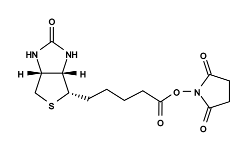
图1. ReadiLink 蛋白质生物素化试剂盒的化学结构


参考文献

A low-cost, high-performance system for fluorescence lateral flow assays
Authors: Linda G Lee, Eric S Nordman, Martin D Johnson, Mark F Oldham
Journal: Biosensors (2013): 360–373

Implementation of P22 viral capsids as nanoplatforms
Authors: Sebyung Kang, Masaki Uchida, Alison O’Neil, Rui Li, Peter E Prevelige, Trevor Douglas
Journal: Biomacromolecules (2010): 2804–2809

Calcium-dependent activation of transglutaminase 2 by nanosecond pulsed electric fields
Authors: Keiko Morotomi-Yano, Ken-ichi Yano
Journal: FEBS Open Bio


相关产品

产品名称 货号
ReadiLink 蛋白质生物素偶联试剂盒 Cat#5521

Amplite 比色法葡萄糖氧化酶检测试剂盒 红色荧光 货号11299-AAT Bioquest荧光染料

上海金畔生物科技有限公司代理AAT Bioquest荧光染料全线产品,欢迎访问AAT Bioquest荧光染料官网了解更多信息。

Amplite 比色法葡萄糖氧化酶检测试剂盒 红色荧光

Amplite 比色法葡萄糖氧化酶检测试剂盒 红色荧光

Amplite 比色法葡萄糖氧化酶检测试剂盒 红色荧光     货号11299 货号 11299 存储条件 在零下15度以下保存, 避免光照
规格 500 Tests 价格 3924
Ex (nm) 571 Em (nm) 584
分子量 溶剂
产品详细介绍

简要概述

Amplite 比色法葡萄糖氧化酶检测试剂盒是美国AAT Bioquest生产的用于定量蛋白的试剂盒,葡萄糖氧化酶是二聚体蛋白质,其催化β-D-葡萄糖氧化成过氧化氢和D-葡糖酸-1,5-内酯,其被水解成葡糖酸。 它广泛用于测定体液中的葡萄糖以及从饮料,食品和其他农产品中去除残留的葡萄糖和氧气。 此外,葡萄糖氧化酶通常用于生物传感器中以检测葡萄糖。 Amplite 葡萄糖氧化酶检测试剂盒为溶液中葡萄糖氧化酶的测量提供了一种快速而灵敏的方法。 它可以以方便的96孔或384孔微量滴定板形式进行,并且很容易适应自动化而无需分离步骤。 该试剂盒使用我们的Amplite Red底物,可使用570 nm的吸光度酶标仪进行监测。 使用Amplite 比色葡萄糖氧化酶检测试剂盒,我们检测到低至12.5 mU / mL的葡萄糖氧化酶。金畔生物是AAT Bioquest的中国代理商,为您提供最优质的Amplite 比色法葡萄糖氧化酶检测试剂盒。 

点击查看光谱


适用仪器


光吸收酶标仪  
吸收: 570nm
推荐孔板: 透明底板

产品说明书

样品实验方案

简要概述

1.准备工作溶液(50μL)
2.添加葡萄糖氧化酶标准品或测试样品(50μL)
3.在37°C孵育10-30分钟
4.监测OD = 570nm处的吸光度


溶液制备 

1.储备溶液

所有未使用的储备溶液应分成一次性等分试样,并在制备后储存在-20°C。避免反复冻融循环。
1.1 Amplite 红色储备液(250X):
将100μLDMSO(组分E)加入到Amplite Red(组分A)的小瓶中。注意:在硫醇如二硫苏糖醇(DTT)和2-巯基乙醇存在下,Amplite Red不稳定。反应中DTT或2-巯基乙醇的最终浓度应不高于10μM。 Amplite Red在高pH(> 8.5)下也不稳定。因此,反应应在pH 7-8下进行。推荐提供的测定缓冲液(pH 7.4)。

1.2 HRP库存解决方案(50X):
将1mL测定缓冲液(组分B)加入到辣根过氧化物酶(组分C)的小瓶中。

1.3 葡萄糖氧化酶原液(100 U / mL):
将1mL测定缓冲液(组分B)加入葡萄糖氧化酶(组分D)的小瓶中。

1.4 葡萄糖原液(10X):
将5mL测定缓冲液(组分B)加入葡萄糖小瓶(组分F)中。


2.标准溶液

葡萄糖氧化酶标准
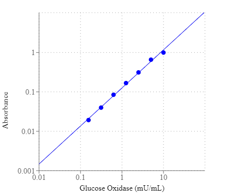
通过将2μL的100U / mL葡萄糖氧化酶储备溶液稀释到200μL测定缓冲液(组分B)中来制备葡萄糖氧化酶标准品,以具有1000mU / mL葡萄糖氧化酶标准溶液。 然后进行1:100连续稀释,然后进行1:2连续稀释,得到连续稀释的葡萄糖氧化酶标准品,从10 mU / mL至0.156 mU / mL(GOS1 – GOS7)。


3.工作溶液

将20μLDelterite Red储备液(250X),100μLHRP储备液(50X)和500μL葡萄糖储备液(10X)加入4.3 mL分析缓冲液(组分B)中,制成5 mL工作溶液。


样品分析

表1.透明底96孔微孔板中葡萄糖氧化酶标准品和测试样品的布局。 GOS =葡萄糖氧化酶标准品(GOS1-GOS7,0.156至10mU / mL),BL =空白对照,TS =测试样品。

BL BL TS TS
GOS1 GOS1
GOS2 GOS2
GOS3 GOS3    
GOS4 GOS4    
GOS5 GOS5    
GOS6 GOS6    
GOS7 GOS7    

表2.每个孔的试剂组成

容积 试剂
GOS1-GOS7 50ul 连续稀释(0.156至10 mU / mL)
BL 50ul 分析缓冲液(组分B)
TS 50ul 测试样本

1.根据表1和2中提供的布局制备葡萄糖氧化酶标准品(GOS),空白对照(BL)和测试样品(TS)。对于384孔板,每孔使用25μL试剂代替50μL。

2.向葡萄糖氧化酶标准品,空白对照和测试样品的每个孔中加入50μL工作溶液,使总测定体积为100μL/孔。 对于384孔板,在每个孔中加入25μLGO工作溶液,总体积为50μL/孔。

3.将反应在37°C孵育10至30分钟,避光。

4.用OD = 570nm的吸光度板读数器监测吸光度的增加。


参考文献

A reassessment of the carnivorous status of salmonids: Hepatic glucokinase is expressed in wild fish in Kerguelen Islands
Authors: Lucie Marandel, Philippe Gaudin, Frančois Guéraud, Stéphane Glise, Alexandre Herman, Elisabeth Plagnes-Juan, Vincent Véron, Stéphane Panserat, Jacques Labonne
Journal: Science of The Total Environment (2018): 276–285

Overcoming 5-Fu resistance in human non-small cell lung cancer cells by the combination of 5-Fu and cisplatin through the inhibition of glucose metabolism
Authors: Jun-gang Zhao, Kai-ming Ren, Jun Tang
Journal: Tumor Biology (2014): 12305–12315


相关产品

产品名称 货号
Amplite 比色法黄嘌呤氧化酶检测试剂盒 Cat#11307
Amplite 荧光法葡萄糖氧化酶检测试剂盒 Cat#11300
Amplite NADPH检测试剂盒(比色法) Cat#15272

Portelite 荧光蛋白定量试剂盒 优化用于 CytoCite 和Qubit 荧光分析仪 货号11109-AAT Bioquest荧光染料

上海金畔生物科技有限公司代理AAT Bioquest荧光染料全线产品,欢迎访问AAT Bioquest荧光染料官网了解更多信息。

Portelite 荧光蛋白定量试剂盒 优化用于 CytoCite 和Qubit 荧光分析仪

Portelite 荧光蛋白定量试剂盒 优化用于 CytoCite 和Qubit 荧光分析仪

Portelite 荧光蛋白定量试剂盒 优化用于 CytoCite 和Qubit 荧光分析仪    货号11109 货号 11109 存储条件 在2-8度冷藏保存
规格 100 Tests 价格 1008
Ex (nm) 485 Em (nm) 590
分子量 溶剂
产品详细介绍

简要概述

Portelite 荧光蛋白定量试剂盒是美国AAT Bioquest生产的用于定量蛋白的试剂盒,蛋白质定量是蛋白质纯化,电泳,细胞生物学,分子生物学和其他研究应用中的重要组成部分。 Biuret,Lowry,BCA和Bradford检测常规用于估计蛋白质浓度。然而,这些比色测定不太敏感,并且需要大的样品体积以确保准确性。我们的Portelite 荧光蛋白定量试剂盒比现有的比色蛋白测定法(例如Bradford和Bicinchoninic acid(BCA)测定法)更敏感。试剂盒中使用的Prolite 橙在水溶液中是非荧光的,但与蛋白质反应迅速并产生明亮的荧光。 Portelite 荧光蛋白定量试剂盒提供了一种简单的方法来定量溶液中的蛋白质浓度。该测定的动态范围为12.5ug / mL至5mg / mL的BSA。该套件针对Cytocite 和Qubit 荧光分析仪进行了优化。它可用于(1)研究蛋白质/蛋白质相互作用; (2)亲和层析后测定柱级分; (3)估计细胞提取物中膜蛋白的回收率; (4)融合蛋白的高通量筛选。金畔生物是AAT Bioquest的中国代理商,为您提供最优质的Portelite 荧光蛋白定量试剂盒。 


适用仪器


Qubit 荧光计  
激发: 480nm
发射: 510-580nm
器材: 0.2 mL薄壁PCR管
CytoCite 荧光计  
激发: 480nm
发射: 510-580nm
器材: 0.2 mL薄壁PCR管

产品说明书

操作步骤

简要概述

准备并添加BSA标准品或测试样品(10μL)
在0.2 mL PCR管(Cat#CCT100)中制备并添加Prolite 橙色工作溶液(190μL)
在室温下孵育15分钟
使用CytoCite 或Qubit荧光分析仪监测荧光


溶液制备

Prolite 橙色工作溶液:
将5μLProlite Orange(200X)(组分A)加入995μL样品稀释缓冲液(组分E)中并充分混合。 注意:请勿将工作溶液混入玻璃容器中。


样品实验方案

该方案适用于Qubit®荧光分析仪。

1.运行蛋白质测定

1.1每孔加入190μL/孔的Prolite Orange工作溶液。

1.2将10μLBSA标准品(组分B,C,D)或10μL样品加入190μLProlite Orange工作溶液管中,使最终测定体积为200μL/管。

1.3在室温下孵育反应15分钟。 注意:保护样品避光,避免将样品拿在手中。

1.4将样品插入CytoCite 并用绿色荧光通道监测荧光。 按照适用于CytoCite 荧光计的步骤进行操作。具体操作点击查看。


2.Qubit®荧光计的简要方案

2.1按Qubit®主屏幕主屏幕上的蛋白质,然后按读取标准。

2.2将3个含有标准的试管中的每一个插入样品室。

2.3关闭盖子并按Read标准。

2.4仪器显示结果并生成校准曲线。

2.5按Run sample并选择样品量至10μL。

2.6将样品管插入样品室。

2.7盖上盖子,然后按Read tube。

2.8仪器在实验屏幕上显示结果。 最高值是原始样品浓度,最低值是稀释浓度。


3.标准溶液制备

        对于CytoCite 荧光计测定,您可以选择使用自己的蛋白质标准进行校准。这是一个生成定制蛋白质标准曲线的简要方案。

3.1在PBS缓冲液中制备400μg/ ml(400 ng /μL)的蛋白质溶液。

3.2用PBS缓冲液进行1:2连续稀释,得到200,100,50,25,12.5ng /μl系列标准稀释液。

3.3将190μLPolite 橙色工作溶液加入0.2 mL PCR管中。

3.4每管加入10μL标准品或10μL样品。

3.5在室温下孵育反应15分钟。

3.6将样品插入CytoCite 并用绿色荧光通道监测荧光。


图示

Portelite 荧光蛋白定量试剂盒 优化用于 CytoCite 和Qubit 荧光分析仪    货号11109

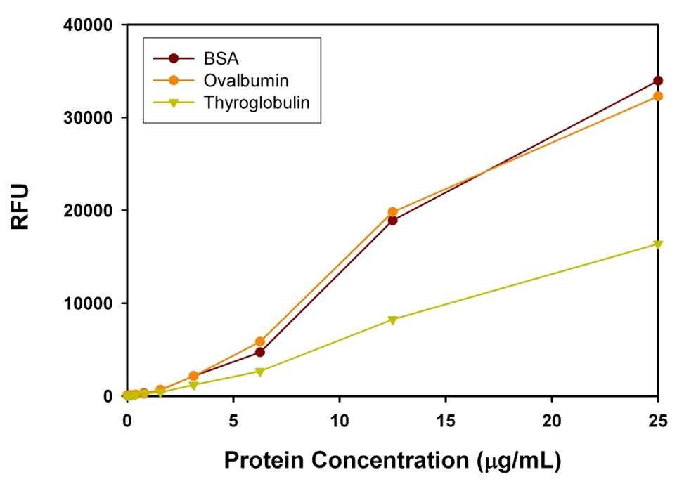
图1使用Portelite 荧光蛋白定量试剂盒*优化用于CytoCite 和Qubit 荧光分析仪*和Qubit®荧光分析仪,在Ex / Em 485 / 5000nm下测量BSA,鸡蛋卵清蛋白,猪甲状腺球蛋白的系列稀释液。 可以检测到低至50 ng / mL的蛋白质。


参考文献

Dual Amplification Fluorescence Assay for Alpha Fetal Protein Utilizing Immunohybridization Chain Reaction and Metal-Enhanced Fluorescence of Carbon Nanodots
Authors: Xu, D. D.; Liu, C.; Li, C. Y.; Song, C. Y.; Kang, Y. F.; Qi, C. B.; Lin, Y.; Pang, D. W.; Tang, H. W.
Journal: ACS Appl Mater Interfaces (2017): 37606-37614

Quantification of Membrane Protein Self-Association with a High-Throughput Compatible Fluorescence Assay
Authors: Li, J.; Qiu, X. J.
Journal: Biochemistry (2017): 1951-1954

Use of anchor protein modules in fluorescence polarisation aptamer assay for ochratoxin A determination
Authors: Samokhvalov, A. V.; Safenkova, I. V.; Eremin, S. A.; Zherdev, A. V.; Dzantiev, B. B.
Journal: Anal Chim Acta (2017): 80-87

Tryptophan fluorescence quenching as a binding assay to monitor protein conformation changes in the membrane of intact mitochondria
Authors: Akbar, S. M.; Sreeramulu, K.; Sharma, H. C.
Journal: J Bioenerg Biomembr (2016): 241-7

Ag@SiO2-entrapped hydrogel microarray: a new platform for a metal-enhanced fluorescence-based protein assay
Authors: Jang, E.; Kim, M.; Koh, W. G.
Journal: Analyst (2015): 3375-83

Label-free fluorescence assay for protein kinase based on peptide biomineralized gold nanoclusters as signal sensing probe
Authors: Song, W.; Wang, Y.; Liang, R. P.; Zhang, L.; Qiu, J. D.
Journal: Biosens Bioelectron (2015): 234-40

Budded baculoviruses as a tool for a homogeneous fluorescence anisotropy-based assay of ligand binding to G protein-coupled receptors: the case of melanocortin 4 receptors
Authors: Veiksina, S.; Kopanchuk, S.; Rinken, A.
Journal: Biochim Biophys Acta (2014): 372-81

Characterization of G protein-coupled receptors by a fluorescence-based calcium mobilization assay
Authors: Caers, J.; Peymen, K.; Suetens, N.; Temmerman, L.; Janssen, T.; Schoofs, L.; Beets, I.
Journal: J Vis Exp (2014): e51516

Cleavage of pro-tumor necrosis factor alpha by ADAM metallopeptidase domain 17: a fluorescence-based protease assay cleaves its natural protein substrate
Authors: Zhang, C.; Zheng, L.; Nurnberg, J.; Vacari, B. M.; Zhou, J.; Wang, Y.
Journal: Anal Biochem (2014): 14-9

A fluorescence-based thermal shift assay identifies inhibitors of mitogen activated protein kinase kinase 4
Authors: Krishna, S. N.; Luan, C. H.; Mishra, R. K.; Xu, L.; Scheidt, K. A.; Anderson, W. F.; Bergan, R. C.
Journal: PLoS One (2013): e81504

Amplite 荧光素酶报告基因检测试剂盒 货号12518-AAT Bioquest荧光染料

上海金畔生物科技有限公司代理AAT Bioquest荧光染料全线产品,欢迎访问AAT Bioquest荧光染料官网了解更多信息。

Amplite 荧光素酶报告基因检测试剂盒

Amplite 荧光素酶报告基因检测试剂盒

Amplite 荧光素酶报告基因检测试剂盒    货号12518 货号 12518 存储条件 在零下15度以下保存, 避免光照
规格 1 plate 价格 1944
Ex (nm) Em (nm)
分子量 溶剂
产品详细介绍

简要概述

Amplite 荧光素酶报告基因检测试剂盒是美国AAT Bioquest生产的用于荧光素酶报告基因检测的试剂盒。β-半乳糖苷酶,β-葡萄糖醛酸酶和荧光素酶是常见的报告基因。最好的多功能常见报告基因是来自于北美的萤火虫Photinus pyralis的荧光素酶。此酶蛋白的活性不需要进行蛋白质翻译后修饰获得。它在活细胞中高浓度积累不会造成细胞毒性,可以在原核和真核细胞中使用。在ATP、镁离子和氧存在的条件下,萤火虫荧光素酶催化荧光素氧化形成生物荧光。Amplite 荧光素酶报告基因检测试剂盒利用与荧光强度相关的公式来定量检测活细胞和细胞抽提物中的荧光素酶。在荧光素酶的催化下,我们的试剂/试剂盒会产生具有强烈荧光的荧光产物。本试剂盒提供所有的必需组分,并且经过优化处理,我们还提供可以用于HTS(高通量筛选)实验方案。本试剂盒具有非常好的灵敏度,可以用于对灵敏度有要求的分析。金畔生物是AAT Bioquest的中国代理商,为您提供最优质的Amplite 荧光素酶报告基因检测试剂盒。 

金畔问号:荧光素酶到底该如何检测?


适用仪器


发光酶标仪  
推荐孔板: 白色孔板

产品说明书

样品实验方案

简要概述

1.准备细胞(样品)(96孔板是100 µL /孔,384孔板是25 µL /孔)
2.添加等量的萤光素酶工作溶液
3.在室温下孵育10-20分钟
4.检测560 nm处的荧光强度


溶液制备 

1.工作溶液配制

将全部反应缓冲液(组分B)转移到荧光素酶(组分A)的瓶中,并充分混合以制成荧光素酶工作溶液。

点击查看细胞制备指南


样品操作及分析

1.进行荧光素酶检测:

1.1通过在所需化合物缓冲液中加入10 µL 10X测试化合物(96孔板)或5 µL 5X测试化合物(384孔板)来用检测化合物处理细胞(或样品)。 对于空白孔(没有细胞的培养基),添加相应量的化合物缓冲液。

1.2将细胞板在5%CO2培养箱中于37°C孵育一段时间(通常为4小时至过夜)。

1.3每孔荧光素酶工作溶液添加100 µL(96孔板)或25 µL(384孔板)。

1.4避光在室温下孵育平板10-20分钟。

1.5用酶标仪检测荧光强度。


2.建立标准的萤光素酶校准曲线:注意:如果需要计算样品中萤光素酶的绝对量,则应与上述测定法一起生成萤光素酶标准曲线。

2.1使用不含萤光素酶的样品(作为对照)来检测背景发光,在含0.1%BSA的PBS缓冲液中进行一系列萤光素酶稀释液。 注意:通常萤光素酶的浓度从1 pg / mL到1 ng / mL是合适的。

2.2将100 µL /孔(96孔板)或25 µL /孔(384孔板)的稀释的荧光素酶溶液添加到一个空板中。

2.3加入100 µL /孔(96孔板)或25 µL /孔(384孔板)的荧光素酶工作溶液。

2.4在避光的条件下,将反应混合物在室温下孵育10-20分钟。

2.5用标准酶标仪记录荧光强度。

2.6生成荧光素酶标准曲线。


参考文献

LncRNA TUBA4B functions as a competitive endogenous RNA to inhibit gastric cancer progression by elevating PTEN via sponging miR-214 and miR-216a/b
Authors: Jianbo Guo, Yan Li, He Duan, Lu Yuan
Journal: Cancer Cell International (2019): 156

Identification of compounds that modulate retinol signaling using a cell-based qHTS assay
Authors: Yanling Chen, Srilatha Sakamuru, Ruili Huang, David H Reese, Menghang Xia
Journal: Toxicology in Vitro (2016): 287–296

Microwave ablation-assisted liver gene transfection in rats
Authors: Ruoyu Jiang, Lingkai Meng, Longhao Sun, Xianghui He, Xiaoyu Liang, Jie Zhang, Zhixiang Zhang
Journal: International Journal of Hyperthermia (2016): 666–672

Activation of relaxin family receptor 1 from different mammalian species by relaxin peptide and small-molecule agonist ML290
Authors: Zaohua Huang, Courtney Myhr, Ross AD Bathgate, Brian A Ho, Amaya Bueno, Xin Hu, Jingbo Xiao, Noel Southall, Elena Barnaeva, Irina U Agoulnik
Journal: Frontiers in endocrinology (2015)

Neuroprotective effect of schizandrin A on oxygen and glucose deprivation/reperfusion-induced cell injury in primary culture of rat cortical neurons
Authors: Cai-Ping Wang, Gui-Cai Li, Yun-Wei Shi, Xiao-Chuan Zhang, Jian-Long Li, Zhi-Wei Wang, Fei Ding, Xin-Miao Liang
Journal: Journal of physiology and biochemistry (2014): 735–747

Discovery of ML367, inhibitor of ATAD5 stabilization
Authors: Jason M Rohde, Ganesha Rai, Yong Jun Choi, Srilatha Sakamuru, Jennifer T Fox, Ruili Huang, Menghang Xia, Kyungjae Myung, Matthew B Boxer, David J Maloney
Journal: (2013)

Amplite 荧光素酶报告基因检测试剂盒 货号12519-AAT Bioquest荧光染料

上海金畔生物科技有限公司代理AAT Bioquest荧光染料全线产品,欢迎访问AAT Bioquest荧光染料官网了解更多信息。

Amplite 荧光素酶报告基因检测试剂盒

Amplite 荧光素酶报告基因检测试剂盒

Amplite 荧光素酶报告基因检测试剂盒    货号12519 货号 12519 存储条件 在零下15度以下保存, 避免光照
规格 10 plates 价格 5244
Ex (nm) Em (nm)
分子量 溶剂
产品详细介绍

简要概述

Amplite 荧光素酶报告基因检测试剂盒是美国AAT Bioquest生产的用于荧光素酶报告基因检测的试剂盒。β-半乳糖苷酶,β-葡萄糖醛酸酶和荧光素酶是常见的报告基因。最好的多功能常见报告基因是来自于北美的萤火虫Photinus pyralis的荧光素酶。此酶蛋白的活性不需要进行蛋白质翻译后修饰获得。它在活细胞中高浓度积累不会造成细胞毒性,可以在原核和真核细胞中使用。在ATP、镁离子和氧存在的条件下,萤火虫荧光素酶催化荧光素氧化形成生物荧光。Amplite 荧光素酶报告基因检测试剂盒利用与荧光强度相关的公式来定量检测活细胞和细胞抽提物中的荧光素酶。在荧光素酶的催化下,我们的试剂/试剂盒会产生具有强烈荧光的荧光产物。本试剂盒提供所有的必需组分,并且经过优化处理,我们还提供可以用于HTS(高通量筛选)实验方案。本试剂盒具有非常好的灵敏度,可以用于对灵敏度有要求的分析。金畔生物是AAT Bioquest的中国代理商,为您提供最优质的Amplite 荧光素酶报告基因检测试剂盒。 

金畔问号:荧光素酶到底该如何检测?


适用仪器


发光酶标仪  
推荐孔板: 白色孔板

产品说明书

样品实验方案

简要概述

1.准备细胞(样品)(96孔板是100 µL /孔,384孔板是25 µL /孔)
2.添加等量的萤光素酶工作溶液
3.在室温下孵育10-20分钟
4.检测560 nm处的荧光强度


溶液制备 

1.工作溶液配制

将全部反应缓冲液(组分B)转移到荧光素酶(组分A)的瓶中,并充分混合以制成荧光素酶工作溶液。

点击查看细胞制备指南


样品操作及分析

1.进行荧光素酶检测:

1.1通过在所需化合物缓冲液中加入10 µL 10X测试化合物(96孔板)或5 µL 5X测试化合物(384孔板)来用检测化合物处理细胞(或样品)。 对于空白孔(没有细胞的培养基),添加相应量的化合物缓冲液。

1.2将细胞板在5%CO2培养箱中于37°C孵育一段时间(通常为4小时至过夜)。

1.3每孔荧光素酶工作溶液添加100 µL(96孔板)或25 µL(384孔板)。

1.4避光在室温下孵育平板10-20分钟。

1.5用酶标仪检测荧光强度。


2.建立标准的萤光素酶校准曲线:注意:如果需要计算样品中萤光素酶的绝对量,则应与上述测定法一起生成萤光素酶标准曲线。

2.1使用不含萤光素酶的样品(作为对照)来检测背景发光,在含0.1%BSA的PBS缓冲液中进行一系列萤光素酶稀释液。 注意:通常萤光素酶的浓度从1 pg / mL到1 ng / mL是合适的。

2.2将100 µL /孔(96孔板)或25 µL /孔(384孔板)的稀释的荧光素酶溶液添加到一个空板中。

2.3加入100 µL /孔(96孔板)或25 µL /孔(384孔板)的荧光素酶工作溶液。

2.4在避光的条件下,将反应混合物在室温下孵育10-20分钟。

2.5用标准酶标仪记录荧光强度。

2.6生成荧光素酶标准曲线。


参考文献

LncRNA TUBA4B functions as a competitive endogenous RNA to inhibit gastric cancer progression by elevating PTEN via sponging miR-214 and miR-216a/b
Authors: Jianbo Guo, Yan Li, He Duan, Lu Yuan
Journal: Cancer Cell International (2019): 156

Identification of compounds that modulate retinol signaling using a cell-based qHTS assay
Authors: Yanling Chen, Srilatha Sakamuru, Ruili Huang, David H Reese, Menghang Xia
Journal: Toxicology in Vitro (2016): 287–296

Microwave ablation-assisted liver gene transfection in rats
Authors: Ruoyu Jiang, Lingkai Meng, Longhao Sun, Xianghui He, Xiaoyu Liang, Jie Zhang, Zhixiang Zhang
Journal: International Journal of Hyperthermia (2016): 666–672

Activation of relaxin family receptor 1 from different mammalian species by relaxin peptide and small-molecule agonist ML290
Authors: Zaohua Huang, Courtney Myhr, Ross AD Bathgate, Brian A Ho, Amaya Bueno, Xin Hu, Jingbo Xiao, Noel Southall, Elena Barnaeva, Irina U Agoulnik
Journal: Frontiers in endocrinology (2015)

Neuroprotective effect of schizandrin A on oxygen and glucose deprivation/reperfusion-induced cell injury in primary culture of rat cortical neurons
Authors: Cai-Ping Wang, Gui-Cai Li, Yun-Wei Shi, Xiao-Chuan Zhang, Jian-Long Li, Zhi-Wei Wang, Fei Ding, Xin-Miao Liang
Journal: Journal of physiology and biochemistry (2014): 735–747

Discovery of ML367, inhibitor of ATAD5 stabilization
Authors: Jason M Rohde, Ganesha Rai, Yong Jun Choi, Srilatha Sakamuru, Jennifer T Fox, Ruili Huang, Menghang Xia, Kyungjae Myung, Matthew B Boxer, David J Maloney
Journal: (2013)

Amplite 荧光素酶报告基因检测试剂盒 货号12520-AAT Bioquest荧光染料

上海金畔生物科技有限公司代理AAT Bioquest荧光染料全线产品,欢迎访问AAT Bioquest荧光染料官网了解更多信息。

Amplite 荧光素酶报告基因检测试剂盒

Amplite 荧光素酶报告基因检测试剂盒

Amplite 荧光素酶报告基因检测试剂盒    货号12520 货号 12520 存储条件 在零下15度以下保存, 避免光照
规格 100 plates 价格 39060
Ex (nm) Em (nm)
分子量 溶剂
产品详细介绍

简要概述

Amplite 荧光素酶报告基因检测试剂盒是美国AAT Bioquest生产的用于荧光素酶报告基因检测的试剂盒。β-半乳糖苷酶,β-葡萄糖醛酸酶和荧光素酶是常见的报告基因。最好的多功能常见报告基因是来自于北美的萤火虫Photinus pyralis的荧光素酶。此酶蛋白的活性不需要进行蛋白质翻译后修饰获得。它在活细胞中高浓度积累不会造成细胞毒性,可以在原核和真核细胞中使用。在ATP、镁离子和氧存在的条件下,萤火虫荧光素酶催化荧光素氧化形成生物荧光。Amplite 荧光素酶报告基因检测试剂盒利用与荧光强度相关的公式来定量检测活细胞和细胞抽提物中的荧光素酶。在荧光素酶的催化下,我们的试剂/试剂盒会产生具有强烈荧光的荧光产物。本试剂盒提供所有的必需组分,并且经过优化处理,我们还提供可以用于HTS(高通量筛选)实验方案。本试剂盒具有非常好的灵敏度,可以用于对灵敏度有要求的分析。金畔生物是AAT Bioquest的中国代理商,为您提供最优质的Amplite 荧光素酶报告基因检测试剂盒。 

金畔问号:荧光素酶到底该如何检测?


适用仪器


发光酶标仪  
推荐孔板: 白色孔板

产品说明书

样品实验方案

简要概述

1.准备细胞(样品)(96孔板是100 µL /孔,384孔板是25 µL /孔)
2.添加等量的萤光素酶工作溶液
3.在室温下孵育10-20分钟
4.检测560 nm处的荧光强度


溶液制备 

1.工作溶液配制

将全部反应缓冲液(组分B)转移到荧光素酶(组分A)的瓶中,并充分混合以制成荧光素酶工作溶液。

点击查看细胞制备指南


样品操作及分析

1.进行荧光素酶检测:

1.1通过在所需化合物缓冲液中加入10 µL 10X测试化合物(96孔板)或5 µL 5X测试化合物(384孔板)来用检测化合物处理细胞(或样品)。 对于空白孔(没有细胞的培养基),添加相应量的化合物缓冲液。

1.2将细胞板在5%CO2培养箱中于37°C孵育一段时间(通常为4小时至过夜)。

1.3每孔荧光素酶工作溶液添加100 µL(96孔板)或25 µL(384孔板)。

1.4避光在室温下孵育平板10-20分钟。

1.5用酶标仪检测荧光强度。


2.建立标准的萤光素酶校准曲线:注意:如果需要计算样品中萤光素酶的绝对量,则应与上述测定法一起生成萤光素酶标准曲线。

2.1使用不含萤光素酶的样品(作为对照)来检测背景发光,在含0.1%BSA的PBS缓冲液中进行一系列萤光素酶稀释液。 注意:通常萤光素酶的浓度从1 pg / mL到1 ng / mL是合适的。

2.2将100 µL /孔(96孔板)或25 µL /孔(384孔板)的稀释的荧光素酶溶液添加到一个空板中。

2.3加入100 µL /孔(96孔板)或25 µL /孔(384孔板)的荧光素酶工作溶液。

2.4在避光的条件下,将反应混合物在室温下孵育10-20分钟。

2.5用标准酶标仪记录荧光强度。

2.6生成荧光素酶标准曲线。


参考文献

LncRNA TUBA4B functions as a competitive endogenous RNA to inhibit gastric cancer progression by elevating PTEN via sponging miR-214 and miR-216a/b
Authors: Jianbo Guo, Yan Li, He Duan, Lu Yuan
Journal: Cancer Cell International (2019): 156

Identification of compounds that modulate retinol signaling using a cell-based qHTS assay
Authors: Yanling Chen, Srilatha Sakamuru, Ruili Huang, David H Reese, Menghang Xia
Journal: Toxicology in Vitro (2016): 287–296

Microwave ablation-assisted liver gene transfection in rats
Authors: Ruoyu Jiang, Lingkai Meng, Longhao Sun, Xianghui He, Xiaoyu Liang, Jie Zhang, Zhixiang Zhang
Journal: International Journal of Hyperthermia (2016): 666–672

Activation of relaxin family receptor 1 from different mammalian species by relaxin peptide and small-molecule agonist ML290
Authors: Zaohua Huang, Courtney Myhr, Ross AD Bathgate, Brian A Ho, Amaya Bueno, Xin Hu, Jingbo Xiao, Noel Southall, Elena Barnaeva, Irina U Agoulnik
Journal: Frontiers in endocrinology (2015)

Neuroprotective effect of schizandrin A on oxygen and glucose deprivation/reperfusion-induced cell injury in primary culture of rat cortical neurons
Authors: Cai-Ping Wang, Gui-Cai Li, Yun-Wei Shi, Xiao-Chuan Zhang, Jian-Long Li, Zhi-Wei Wang, Fei Ding, Xin-Miao Liang
Journal: Journal of physiology and biochemistry (2014): 735–747

Discovery of ML367, inhibitor of ATAD5 stabilization
Authors: Jason M Rohde, Ganesha Rai, Yong Jun Choi, Srilatha Sakamuru, Jennifer T Fox, Ruili Huang, Menghang Xia, Kyungjae Myung, Matthew B Boxer, David J Maloney
Journal: (2013)

Amplite NAD/NADH比率检测试剂盒(比色法) 货号15273-AAT Bioquest荧光染料

上海金畔生物科技有限公司代理AAT Bioquest荧光染料全线产品,欢迎访问AAT Bioquest荧光染料官网了解更多信息。

Amplite NAD/NADH比率检测试剂盒(比色法)

Amplite NAD/NADH比率检测试剂盒(比色法)

Amplite NAD/NADH比率检测试剂盒(比色法)    货号15273 货号 15273 存储条件 在零下15度以下保存, 避免光照
规格 250 Tests 价格 4584
Ex (nm) 460 Em (nm)
分子量 溶剂
产品详细介绍

简要概述

Amplite NAD/NADH比率检测试剂盒(比色法)是美国AAT Bioquest研发的检测NAD/NADH的试剂盒,烟酰胺腺嘌呤二核苷酸(NAD)和烟酰胺腺嘌呤二核苷酸磷酸(NADP)是细胞中发现的两种重要的辅酶。 NAD形成NADP,通过酯键将磷酸基团添加到腺苷核苷酸的2’位置。 NADH(NADPH)是NAD(NADP)的还原形式,NAD(NADP)是NADH(NADPH)的氧化形式。 NAD或NADP在氧化还原反应中起辅助因子的作用,在细胞反应中转移电子。 氧化形式和还原形式之间的平衡是NAD / NADH(NADP / NADPH)比率。 该比率是指示细胞氧化还原状态的重要组分,并且它是反映代谢活性和细胞健康的测量。 在健康的哺乳动物组织中,游离NAD +和NADH之间的比率的估计可以高达700.相反,NADP / NADPH比率通常为约0.005,因此NADPH是该辅酶的主要形式。

该Amplite 比色NAD / NADH比率分析试剂盒提供了一种比色法,用于测量培养细胞中细胞内总NAD / NADH量和NAD / NADH比率。 在该测定中,裂解物中的NAD可以用NAD提取溶液提取并通过酶反应转化为NADH,然后通过NADH探针识别,在反应后得到黄色染料,其在460nm处具有吸光度。 产生的染料量与细胞裂解物中NAD或NADH的浓度成正比,可用作细胞NAD / NADH浓度的指示剂。金畔生物是AAT Bioquest的中国代理商,为您提供最优质的NAD/NADH检测试剂盒。 

Amplite NAD/NADH比率检测试剂盒(比色法)    货号15273


适用仪器


光吸收酶标仪  
吸收: 460nm
推荐孔板: 透明底板

产品说明书

96孔板检测示例

概述

准备25μLNADH标准品和/或测试样品在室温下

加入25μLNAD提取液孵育15分钟

加入25μL中和溶液加入75μLNAD/ NADH反应混合物在

室温下孵育15分钟至2小时

监测 在460nm处的吸光度

注意:在开始实验之前,在室温下解冻每个试剂盒组分中的一个。


操作步骤

1.准备NADH储备溶液:

将200μLPBS缓冲液加入到NADH标准品(组分C)的小瓶中,得到1mM(1nmol / L)NADH储备溶液。

注意:未使用的NADH储备溶液应分成单次使用的等分试样并储存在-20℃。


2.准备NAD / NADH反应混合物:

2.1将8 mL NADH探针缓冲液(组分B-II)加入到NAD / NADH回收酶混合物(组分A)的瓶中,并充分混合。

2.2将2 mL NADH探针(组分B-I)加入上述瓶中(来自步骤2.1)并充分混合。

注意:这种NAD / NADH反应混合物足以进行125~200次分析。 未使用的NAD / NADH反应混合物应分成一次性等分试样并储存在-20℃。


3.准备连续稀释的NADH标准品(0至10μM):

3.1将10μL1mMNADH储备溶液(来自步骤1)加入990L PBS缓冲液(pH7.4)中,得到10μM(10pmol / L)NADH标准溶液。

注意:稀释的NADH标准溶液不稳定,应在4小时内使用。

3.2取200μL10M NADH标准溶液(来自步骤3.1)进行1:2连续稀释,得到5,2.5,1.25,0.625,0.313,0.156,0.078和0 M系列稀释的NADH标准品。


4.运行总NAD / NADH分析(总共400个分析/试剂盒):

4.1如表1和2中所述,将含有NADH标准品和含NAD / NADH的测试样品的系列稀释液加入白色/透明底96孔微孔板中。

注意:根据需要准备细胞或组织样本NAD / NADH裂解缓冲液(组分G)可用于裂解细胞(详见附录)。


表1.白色/透明底96孔微孔板中NADH标准品和测试样品的布局

BL

BL

TS

TS

NS1

NS1

….

….

NS2

NS2


NS3

NS3


NS4

NS4


NS5

NS5


NS6

NS6


NS7

NS7


注意:NS = NADH标准,BL =空白对照,TS =测试样品


表2.每个孔的试剂组成

NADH Standard

Blank Control

Test Sample

Serial Dilutions*: 50 μL

PBS: 50 μL

50 μL 

*注意:将连续稀释的NADH标准品(0.078μM至5μM)加入NS1至NS7的孔中,一式两份。 高浓度的NADH(例如,>100μM,最终浓度)将导致饱和信号并使校准曲线非线性。


4.2将50μLNAD/ NADH反应混合物(来自步骤2.2)加入NADH标准品,空白对照和测试样品(来自步骤4.1)的每个孔中,以使总NAD / NADH测定体积为100μL/孔。

4.3在室温下孵育反应15分钟至2小时,避光。

4.4使用吸光度读板仪在460 nm处检测吸光度的增加。


5.运行NAD / NADH比率分析(总共250个分析/试剂盒):

5.1如表3和4中所述,将连续稀释的NADH标准品和/或含NAD / NADH的测试样品加入白色/透明96孔微孔板中。

注意:根据需要准备细胞或组织样本。 为方便起见,裂解缓冲液(组分G)可用于裂解细胞(详见附录)。


表3.白色/透明96孔微孔板中NADH标准品和测试样品的布局

BL

BL

TS

TS

TS (NAD)

TS (NAD)

NS1

NS1

….

….

….

….

NS2

NS2


NS3

NS3


NS4

NS4


NS5

NS5


NS6

NS6


NS7

NS7


注意:NS = NAD / NADH标准; BL =空白对照; TS =测试样品; TS(NAD)=用NAD提取溶液(组分D)处理10至15分钟的测试样品,然后用中和溶液(组分E)中和。


表4.每个孔的试剂组合物

NADH Standard

Blank Control

Test Sample (NAD/NADH)

Test Sample (NAD Extract)

Serial Dilutions*: 25 μL

PBS: 25 μL

Test Sample: 25 μL

Test Sample: 25 μL

Component F: 25 μL

Component F: 25 μL

Component F: 25 μL

Component D: 25 μL

Incubate at room temperature for 10 to 15 minutes

Component F: 25 μL

Component F: 25 μL

Component F: 25 μL

Component E: 25 μL

Total: 75 μL

Total: 75 μL

Total: 75 μL

Total: 75 μL

*注意:将连续稀释的NADH标准品(0.078μM至5μM)加入NS1至NS7的孔中,一式两份。 高浓度的NADH(例如,>100μM,最终浓度)将导致饱和信号并使校准曲线非线性。


5.2对于NAD提取(NAD量):将25μLNAD提取溶液(组分D)加入含有NAD / NADH的测试样品的孔中。在室温下孵育10至15分钟,然后加入25μLNADH中和溶液(组分E)以中和NAD提取物,如表3和4中所述。

对于总NAD和NADH(总量):将25μLNAD/ NADH对照溶液(组分F)加入NADH标准品和含有NAD / NADH的测试样品的孔中。在室温下孵育10至15分钟,然后如表3和4中所述添加25μL提取对照溶液(组分F)。

注意:根据需要准备细胞或组织样本。裂解缓冲液(组分G)可用于裂解细胞(详见附录)。

5.3将75μL的NADH反应混合物(来自步骤2.2)加入NADH标准品,空白对照和测试样品(NAD / NADH和NAD提取物)(来自步骤5.1)的每个孔中,使总NADH测定体积为150μL /好。

5.4在室温下孵育反应15分钟至2小时,避光。

5.5使用吸光度读板仪在460 nm处检测吸光度的增加。


数据分析

        空白孔(仅PBS缓冲液)中的吸光度用作对照,并从具有NADH反应的那些孔的值中减去。

Amplite NAD/NADH比率检测试剂盒(比色法)    货号15273

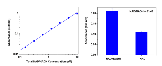
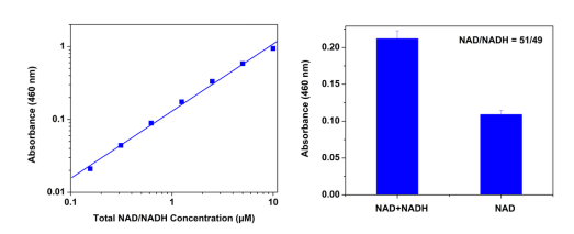
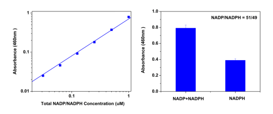
图1. Amplite™比色NAD / NADH比率分析试剂盒用于使用SpectraMax酶标仪(Molecular devices)测量白色/透明96孔微孔板中的总NAD / NADH量和NAD / NADH比率。

A-总NADH和NAD剂量反应:孵育1小时可检测到低至0.1μM的总NADH。

B-NAD / NADH比:在有或没有NAD提取溶液的情况下处理等量的NAD和NADH混合物15分钟,然后在室温下用提取溶液中和。 在460nm读取信号。 基于图1B中所示的吸光度计算NAD / NADH摩尔比。


附录:使用组分D(裂解缓冲液)的测试样品制剂

1.植物细胞样品:用200mg / mL的裂解缓冲液均质化,并以2500rpm离心5-10分钟,使用上清液进行测试。

2.细菌细胞样品:离心收集细菌细胞((10,000 g,0°C,15 min)。使用约1亿至1千万细胞/ mL裂解缓冲液,将处理后的溶液在室温下保持15分钟.2500℃离心 转速5分钟,并用上清液进行试验。

3.哺乳动物细胞样品:从平板孔中取出培养基,每1-5百万个细胞使用约100μL裂解缓冲液(或在96孔细胞培养板中使用50-100μL/孔),并将处理过的溶液保持在室温下 15分钟。 直接使用细胞裂解液或以1500 rpm离心5分钟,使用上清液进行测试。

4.组织样品:称取约20mg组织,用冷PBS洗涤。 在微量离心管中用400μl裂解缓冲液均化。 以2500rpm离心5-10分钟,使用上清液进行测定。


参考文献

β-Lapachone protects against doxorubicin-induced nephrotoxicity via NAD+/AMPK/NF-kB in mice
Authors: Davoud Sanajou, Saeed Nazari Soltan Ahmad, Vahid Hosseini, Ashkan Kalantary-Charvadeh, Yasser Marandi, Leila Roshangar, Saman Bahrambeigi, Mehran Mesgari-Abbasi
Journal: Naunyn-Schmiedeberg’s archives of pharmacology (2019): 1–8

Enhanced NADH Metabolism Involves Colistin-Induced Killing of Bacillus subtilis and Paenibacillus polymyxa
Authors: Zhiliang Yu, Yuyi Zhu, Jianv Fu, Juanping Qiu, Jianhua Yin
Journal: Molecules (2019): 387

Engineering Corynebacterium crenatum for enhancing succinic acid production
Authors: Xiaoju Chen, Yaojie Zhou, Di Zhang
Journal: Journal of Food Biochemistry (2018): e12645

Influence of Boric Acid on Energy Metabolism and Stress Tolerance of Candida albicans
Authors: Martin Schmidt, Dominic Tran-Nguyen, Patrick Chizek
Journal: Journal of Trace Elements in Medicine and Biology (2018)

Loss of sirtuin 1 and mitofusin 2 contributes to enhanced ischemia/reperfusion injury in aged livers
Authors: Sung Kook Chun, Sooyeon Lee, Joseph Flores-Toro, Rebecca Y U, Ming-Jim Yang, Kristina L Go, Thomas G Biel, Catherine E Miney, Schiley Pierre Louis, Brian K Law
Journal: Aging Cell (2018): e12761

Norisoboldine, a natural AhR agonist, promotes Treg differentiation and attenuates colitis via targeting glycolysis and subsequent NAD+/SIRT1/SUV39H1/H3K9me3 signaling pathway
Authors: Qi Lv, Kai Wang, Simiao Qiao, Ling Yang, Yirong Xin, Yue Dai, Zhifeng Wei
Journal: Cell death & disease (2018): 258

Optically-controlled bacterial metabolite for cancer therapy
Authors: Di-Wei Zheng, Ying Chen, Zi-Hao Li, Lu Xu, Chu-Xin Li, Bin Li, Jin-Xuan Fan, Si-Xue Cheng, Xian-Zheng Zhang
Journal: Nature communications (2018)

Stochastic expression of lactate dehydrogenase A induces Escherichia coli persister formation
Authors: Naoki Yamamoto, Rino Isshiki, Yuto Kawai, Daiki Tanaka, Tetsushi Sekiguchi, Shinya Matsumoto, Satoshi Tsuneda
Journal: Journal of Bioscience and Bioengineering (2018)

Celastrol attenuates angiotensin II mediated human umbilical vein endothelial cells damage through activation of Nrf2/ERK1/2/Nox2 signal pathway
Authors: Miao Li, Xin Liu, Yongpeng He, Qingyin Zheng, Min Wang, Yu Wu, Yuanpeng Zhang, Chaoyun Wang
Journal: European Journal of Pharmacology (2017): 124–133

Cytosolic Redox Status of Wine Yeast (Saccharomyces Cerevisiae) under Hyperosmotic Stress during Icewine Fermentation
Authors: Fei Yang, Caitlin Heit, Debra L Inglis
Journal: Fermentation (2017): 61


相关产品

产品名称 货号
Amplite NADP/NADPH比率检测试剂盒(比色法) Cat#15274
Amplite NAD/NADH比率检测试剂盒(荧光法) 红色荧光 Cat#15263
Amplite NADH检测试剂盒(荧光法) 红色荧光 Cat#15261

Amplite NADP/NADPH比率检测试剂盒(比色法) 货号15274-AAT Bioquest荧光染料

上海金畔生物科技有限公司代理AAT Bioquest荧光染料全线产品,欢迎访问AAT Bioquest荧光染料官网了解更多信息。

Amplite NADP/NADPH比率检测试剂盒(比色法)

Amplite NADP/NADPH比率检测试剂盒(比色法)

Amplite NADP/NADPH比率检测试剂盒(比色法)    货号15274 货号 15274 存储条件 在零下15度以下保存, 避免光照
规格 250 Tests 价格 4584
Ex (nm) 460 Em (nm)
分子量 溶剂
产品详细介绍

简要概述

Amplite NADP/NADPH比率检测试剂盒 (比色法)是美国AAT Bioquest研发的检测NADP/NADPH的试剂盒,烟酰胺腺嘌呤二核苷酸(NAD)和烟酰胺腺嘌呤二核苷酸磷酸(NADP)是细胞中发现的两种重要的辅酶。 NAD形成NADP,通过酯键将磷酸基团添加到腺苷核苷酸的2’位置。 NADH(NADPH)是NAD(NADP)的还原形式,NAD(NADP)是NADH(NADPH)的氧化形式。 NAD或NADP在氧化还原反应中起辅助因子的作用,在细胞反应中转移电子。 氧化形式和还原形式之间的平衡是NAD / NADH(NADP / NADPH)比率。 该比率是指示细胞氧化还原状态的重要组分,并且它是反映代谢活性和细胞健康的测量。 在健康的哺乳动物组织中,游离NAD和NADH之间的比率的估计可以高达700。相反,NADP / NADPH比率通常为约0.005,因此NADPH是该辅酶的主要形式。

该Amplite 比色NADP / NADPH比率分析试剂盒提供了一种比色法,用于测量培养细胞中细胞内总NADP / NADPH量和NADP / NADPH比率。 在该测定中,裂解物中的NADPH可以用NADPH提取溶液提取,然后通过NADPH探针识别,在反应后得到黄色染料,其在460nm处具有吸光度。 产生的染料量与细胞裂解物中NADP或NADPH的浓度成正比,可用作细胞NADP / NADPH浓度的指示剂。金畔生物是AAT Bioquest的中国代理商,为您提供最优质的NADP/NADPH检测试剂盒。 

Amplite NADP/NADPH比率检测试剂盒(比色法)    货号15274


适用仪器


光吸收酶标仪  
吸收: 460nm
推荐孔板: 透明底板

产品说明书

96孔板检测示例

概述

准备25μLNADPH标准品和/或测试样品

加入25μLNADPH提取液

在室温下孵育15分钟

加入25μL中和溶液

加入75μLNADPP/ NADPH反应混合物在室温下孵育15分钟至2小时监测 在460nm处的吸光度

注意:在开始实验之前,在室温下解冻每个试剂盒组分中的一个。


操作步骤

1.准备NADPH原液:

将200μLPBS缓冲液加入到NADPH标准品(组分C)的小瓶中,得到1mM(1nmol / L)NADPH储备溶液。

注意:未使用的NADPH原液应分成单次使用的等分试样并储存在-20℃。


2.制备NADP / NADPH反应混合物:

2.1将8mL NADPH探针缓冲液(组分B-II)加入到NADP / NADPH回收酶混合物(组分A)的瓶中,并充分混合。

2.2将2 mL NADPH探针(组分B-I)加入上述瓶中(来自步骤2.1)并充分混合。

注意:这种NADP / NADPH反应混合物足以进行125~200次分析。 未使用的NADP / NADPH反应混合物应分成一次性等分试样并储存在-20℃。


3.准备连续稀释的NADPH标准品(0至2μM):

3.1将2L 1mM NADPH储备溶液(来自步骤1)加入998L PBS缓冲液(pH7.4)中,产生2M(2 pmols / L)NADPH标准溶液。

注意:稀释的NADPH标准溶液不稳定,应在4小时内使用。

3.2取200μL2MNADPH标准溶液(来自步骤3.1)进行1:2连续稀释,得到1,0.5,0.25,0.125,0.0625,0.0313和0M系列稀释的NADPH标准品。


4.运行总NADP / NADPH分析(总共400个分析/试剂盒):

4.1如说明书中的表1和2中所述,将NADPH标准品和含有NADP / NADPH的测试样品的系列稀释液加入白色/透明底96孔微孔板中。

注意:根据需要准备细胞或组织样本。 为方便起见,裂解缓冲液(组分G)可用于裂解细胞。 (详见附录)。

4.2将50μL的NADP / NADPH反应混合物(来自步骤2.2)加入NADPH标准品,空白对照和测试样品(来自步骤4.1)的每个孔中,以使总NADP / NADPH测定体积为100μL/孔。

4.3在室温下孵育反应15分钟至2小时,避光。

4.4使用吸光度读板仪在460 nm处检测吸光度的增加。


5.运行NADP / NADPH比率分析(总共250个分析/试剂盒):

5.1如说明书中的表3和4中所述,将连续稀释的NADPH标准品和/或含NADP / NADPH的测试样品加入白色/透明96孔微量培养板中。

注意:根据需要准备细胞或组织样本。 为方便起见,裂解缓冲液(组分G)可用于裂解细胞。

5.2对于NADPH提取(NADPH量):将25μLNADPH提取溶液(组分D)加入含有NADP / NADPH的测试样品的孔中。在室温下孵育10至15分钟,然后加入25μL中和溶液(组分E)以中和NADPH提取物,如说明书中的表3和4中所述。

对于总NADP和NADPH(总量):将25μLNADPP/ NADPH对照溶液(组分F)加入NADPH标准品的孔和含有NADP / NADPH的测试样品中。在室温下孵育10至15分钟,然后如说明书中的表3和4中所述添加25μL提取对照溶液(组分F)。

注意:根据需要准备细胞或组织样本。为方便起见,裂解缓冲液(组分G)可用于裂解细胞(详见附录)。


5.3将75μL的NADP / NADPH反应混合物(来自步骤2.2)加入NADPH标准品,空白对照和测试样品(NADP / NADPH)的每个孔中,并测试样品(NADPH提取物)(来自步骤5.1)以使总量达到测定体积为150μL/孔。

5.4在室温下孵育反应15分钟至2小时,避光。

5.5使用吸光度读板仪在460 nm处检测吸光度的增加。


数据分析

        空白孔中的吸光度(仅PBS缓冲液)用作对照,并从具有NADPH反应的那些孔的值中减去。

Amplite NADP/NADPH比率检测试剂盒(比色法)    货号15274

图1. Amplite™比色NADP / NADPH比率分析试剂盒用于使用SpectraMax酶标仪(Molecular devices)测量白色/透明96孔微量培养板中的总NADP / NADPH量和NADP / NADPH比率。

A-总NADPH和NADP剂量反应:孵育1小时可检测到低至0.03μM的总NADPH。

B-NADP / NADPH比例:用或不用NADP提取溶液处理等量的NADP和NADPH混合物15分钟,然后在室温下用提取溶液中和。 在460nm读取信号。 NADP / NADPH摩尔比基于图1B中所示的吸光度计算。


附录:使用组分G(NAD / NADH裂解缓冲液)的测试样品制剂

1.植物细胞样品:

用裂解缓冲液以200mg / mL均化,并以2500rpm离心5-10分钟,使用上清液进行测试。


2.细菌样品:

离心收集细菌细胞((10,000 g,0°C,15 min)。使用约1亿至1千万细胞/ mL裂解缓冲液,将处理后的溶液在室温下保持15分钟。以2500 rpm离心5分钟, 用上清液进行试验。


3.哺乳动物细胞样本:

从平板孔中取出培养基,每1-5百万个细胞使用约100μL裂解缓冲液(或在96孔细胞培养板中使用50-100μL/孔),并将处理过的溶液在室温下保持15分钟。 直接使用细胞裂解液或以1500 rpm离心5分钟,使用上清液进行测试。


4.组织样品:

称取约20mg组织,用冷PBS洗涤。 在微量离心管中用400μl裂解缓冲液均化。 以2500rpm离心5-10分钟,使用上清液进行测定。


试剂应用文献

Impact of Genetic Reduction of NMNAT2 on Chemotherapy-Induced Losses in Cell Viability In Vitro and Peripheral Neuropathy In Vivo
Authors: 
Slivicki, Richard A and Ali, Yousuf O and Lu, Hui-Chen and Hohmann, Andrea G
Journal: 
PloS one (2016): e0147620

NMNAT2: HSP90 Complex Mediates Proteostasis in Proteinopathies
Authors: Ali, Yousuf O and Allen, Hunter M and Yu, Lei and Li-Kroeger, David and Bakhshizadehmahmoudi, Dena and Hatcher, Asante and McCabe, Cristin and Xu, Jishu and Bjorklund, Nicole and Taglialatela, Giulio and others
Journal: PLoS Biol (2016): e1002472

ACLY and ACC1 Regulate Hypoxia-Induced Apoptosis by Modulating ETV4 via α-ketoglutarate
Authors: Keenan, Melissa M and Liu, Beiyu and Tang, Xiaohu and Wu, Jianli and Cyr, Derek and Stevens, Robert D and Ilkayeva, Olga and Huang, Zhiqing and Tollini, Laura A and Murphy, Susan K and others
Journal: PLoS Genet (2015): e1005599

Analysis of the nicotinamide phosphoribosyltransferase family provides insight into vertebrate adaptation to different oxygen levels during the water-to-land transition
Authors: Fang, Chengchi and Guan, Lihong and Zhong, Zaixuan and Gan, Xiaoni and He, Shunping
Journal: FEBS journal (2015): 2858–2878

Metformin-induced energy deficiency leads to the inhibition of lipogenesis in prostate cancer cells
Authors: Loubière, Camille and Goiran, Thomas and Laurent, Kathiane and Djabari, Zied and Tanti, Jean-Frančois and Bost, Frédéric
Journal: Oncotarget (2015): 15652

Prediction of intracellular metabolic states from extracellular metabolomic data
Authors: Aurich, Maike K and Paglia, Giuseppe and Rolfsson, Ottar and Hrafnsdóttir, Sigrún and Magnúsdóttir, Manuela and Stefaniak, Magdalena M and Palsson, Bernhard O and Fleming, Ronan MT and Thiele, Ines
Journal: Metabolomics (2015): 603–619


参考文献

Enhanced metabolic activities for ATP production and elevated metabolic flux via pentose phosphate pathway contribute for better CIK cells expansion
Authors: Weiwei Zhang, Huimin Huang, Haibo Cai, Wen-Song Tan
Journal: Cell proliferation (2019)

Soluble α-Klotho treatment protects adenine-induced uremia rats from sciatic nerve damage
Authors: Yingdan Zhao, Jun Ma, Bo Gu, Yang Yi, Hanqing Wang, Zhiyong Guo
Journal: Biomedical Research (2018)

Celastrol attenuates angiotensin II mediated human umbilical vein endothelial cells damage through activation of Nrf2/ERK1/2/Nox2 signal pathway
Authors: Miao Li, Xin Liu, Yongpeng He, Qingyin Zheng, Min Wang, Yu Wu, Yuanpeng Zhang, Chaoyun Wang
Journal: European Journal of Pharmacology (2017): 124–133

Cytosolic Redox Status of Wine Yeast (Saccharomyces Cerevisiae) under Hyperosmotic Stress during Icewine Fermentation
Authors: Fei Yang, Caitlin Heit, Debra L Inglis
Journal: Fermentation (2017): 61

Epigenetic regulation of Runx2 transcription and osteoblast differentiation by nicotinamide phosphoribosyltransferase
Authors: Min Ling, Peixin Huang, Shamima Islam, Daniel P Heruth, Xuanan Li, Li Qin Zhang, Ding-You Li, Zhaohui Hu, Shui Qing Ye
Journal: Cell & Bioscience (2017): 27

MCU-dependent mitochondrial Ca2+ inhibits NAD+/SIRT3/SOD2 pathway to promote ROS production and metastasis of HCC cells
Authors: T Ren, H Zhang, J Wang, J Zhu, M Jin, Y Wu, X Guo, L Ji, Q Huang, H Yang
Journal: Oncogene (2017)

Metabolic and molecular insights into an essential role of nicotinamide phosphoribosyltransferase
Authors: Li Q Zhang, Leon Van Haandel, Min Xiong, Peixin Huang, Daniel P Heruth, Charlie Bi, Roger Gaedigk, Xun Jiang, Ding-You Li, Gerald Wyckoff
Journal: Cell Death & Disease (2017): e2705

Pyrroloquinoline Quinone, a Redox-active o-Quinone, Stimulates Mitochondrial Biogenesis by Activating SIRT1/PGC-1α Signaling Pathway
Authors: Kazuhiro Saihara, Ryosuke Kamikubo, Kazuto Ikemoto, Koji Uchida, Mitsugu Akagawa
Journal: Biochemistry (2017)

Resveratrol attenuates excessive ethanol exposure induced insulin resistance in rats via improving NAD+/NADH ratio
Authors: Gang Luo, Bingqing Huang, Xiang Qiu, Lin Xiao, Ning Wang, Qin Gao, Wei Yang, Liping Hao
Journal: Molecular Nutrition & Food Research (2017)

A Snapshot of the Plant Glycated Proteome STRUCTURAL, FUNCTIONAL, AND MECHANISTIC ASPECTS
Authors: Tatiana Bilova, Elena Lukasheva, Dominic Brauch, Uta Greifenhagen, Gagan Paudel, Elena Tarakhovskaya, Nadezhda Frolova, Juliane Mittasch, Gerd Ulrich Balcke, Alain Tissier
Journal: Journal of Biological Chemistry (2016): 7621–7636


相关产品

产品名称 货号
Amplite NAD/NADH比率检测试剂盒(比色法) Cat#15273
Amplite NADP/NADPH比率检测试剂盒(荧光法) 红色荧光 Cat#15264
Amplite NADPH检测试剂盒(比色法) Cat#15272

Amplite NADP+/NADPH检测试剂盒(比色法)增强灵敏度 货号15276-AAT Bioquest荧光染料

上海金畔生物科技有限公司代理AAT Bioquest荧光染料全线产品,欢迎访问AAT Bioquest荧光染料官网了解更多信息。

Amplite NADP+/NADPH检测试剂盒(比色法)增强灵敏度

Amplite NADP+/NADPH检测试剂盒(比色法)增强灵敏度

Amplite NADP+/NADPH检测试剂盒(比色法)增强灵敏度    货号15276 货号 15276 存储条件 在零下15度以下保存, 避免光照
规格 400 Tests 价格 3924
Ex (nm) 460 Em (nm)
分子量 溶剂
产品详细介绍

简要概述

Amplite 总NADP和NADPH检测试剂盒 (比色法)增强灵敏度是美国AAT Bioquest研发的检测总NADP和NADPH的试剂盒,烟酰胺腺嘌呤二核苷酸(NAD +)和烟酰胺腺嘌呤二核苷酸磷酸(NADP +)是细胞中发现的两种重要的辅因子。 NADH是NAD +的还原形式。 NAD形成NADP,通过酯键将磷酸基团添加到腺苷核苷酸的2’位置。传统的NAD / NADH和NADP / NADPH测定基于监测340nm处NADH或NADPH吸收的变化。 NAD / NADH和NADP / NADPH测定的短紫外波长使得传统方法具有低灵敏度和高干扰。 AAT Bioquest的Amplite™比色总NADP / NADPH检测试剂盒为检测总NADP和NADPH提供了一种便捷的方法。系统中的酶特异性识别酶循环反应中的NADP / NADPH。无需从样品混合物中纯化NADP / NADPH。酶循环反应显着提高了检测灵敏度。 NADPH探针是一种生色传感器,在NADP减少时具有460nm的最大吸光度。 NADPH探针的吸收与溶液中NADPH的浓度成正比。 Amplite™比色总NADP和NADPH分析试剂盒提供灵敏的分析,可在100μL分析体积中检测低至0.03μM的总NADP / NADPH。与试剂盒#15260相比,该试剂盒具有更高的灵敏度。金畔生物是AAT Bioquest的中国代理商,为您提供最优质的总NADP和NADPH检测试剂盒。 


适用仪器


光吸收酶标仪  
吸收: 460nm
推荐孔板: 透明底板

产品说明书

96孔板检测示例

概述

准备NADP / NADPH反应混合物(50μL)

加入NADPH标准品或测试样品(50μL)

在室温下孵育15分钟至2小时

监测460nm处的吸光度

注意:在开始实验之前,在室温下解冻每个试剂盒组分中的一个。


操作步骤

1.准备NADPH原液:

将200μLPBS缓冲液加入到NADPH标准品(组分C)的小瓶中以制备1mM(1nmol / L)NADPH储备溶液。

注意:未使用的NADPH原液应分成单次使用的等分试样并储存在-20℃。


2.制备NADP / NADPH反应混合物:

2.1将8mL NADPH探针缓冲液(组分B-II)加入到NADP / NADPH回收酶混合物(组分A)的瓶中,并充分混合。

2.2将2 mL NADPH探针(组分B-I)加入上述瓶中(来自步骤2.1)并充分混合。

注意:这种NADP / NADPH反应混合物足以进行200次分析。未使用的NADP / NADPH反应混合物应分成一次性等分试样并储存在-20℃。


3.准备NADPH标准品的连续稀释液(0至2μM):

3.1将2L 1mM NADPH储备溶液(来自步骤1)加入998L PBS缓冲液(pH7.4)中,产生2M(2 pmols / L)NADPH标准溶液。

注意:稀释的NADPH标准溶液不稳定,应在4小时内使用。

3.2取200μL2MNADPH标准溶液(来自步骤3.1)进行1:2连续稀释,得到1,0.5,0.25,0.125,0.0625,0.0313和0M系列稀释的NADPH标准品。

3.3如表1和2中所述,将含有NADPH标准品和含NADP / NADPH的测试样品的系列稀释液加入白色/透明底96孔微量培养板中。

注意:根据需要准备细胞或组织样本。为方便起见,裂解缓冲液(组分D)可用于裂解细胞。 (详见附录)。 


表1:白色/透明底96孔微孔板中NADPH标准品和测试样品的布局

BL

BL

TS

TS

NS1

NS1

….

….

NS2

NS2


NS3

NS3


NS4

NS4


NS5

NS5


NS6

NS6


NS7

NS7


注意:NS = NADPH标准,BL =空白对照,TS =测试样品


表2:每个孔的试剂组成

NADPH Standard

Blank Control

Test Sample

Serial Dilutions*: 50 μL

PBS: 50 μL

50 μL 

*注意:将连续稀释的NADPH标准品(0.0313μM至2μM)加入NS1至NS7的孔中,一式两份。 高浓度的NADPH(例如,>30μM,最终浓度)将导致饱和信号并使校准曲线非线性。


4.在上清液反应中运行NADPH测定:

4.1将50μL的NADP / NADPH反应混合物(来自步骤2.2)加入NADPH标准品,空白对照和测试样品的每个孔中(参见步骤3.3),使总NADP / NADPH测定体积为100μL/孔

注意1:对于384孔板,每孔加入25μL样品和25μLNADP/ NADPH反应混合物。

注意2:根据需要准备细胞或组织样本。 为方便起见,裂解缓冲液(组分D)可用于裂解细胞。 (详见附录)。

4.2在室温下孵育反应15分钟至2小时,避光。

4.3用吸光度读板仪在460 nm处检测吸光度的增加。


数据分析

        空白孔中的吸光度(仅PBS缓冲液)用作对照,并从具有NADPH反应的那些孔的值中减去。

Amplite NADP+/NADPH检测试剂盒(比色法)增强灵敏度    货号15276

图1 使用SpectraMax酶标仪(Molecular devices),在96孔白色/透明底板中用Amplite ™比色总NADP和NADPH测定试剂盒*增强灵敏度*测量NADPH剂量反应。孵育1小时(n = 3)可以检测到低至0.03μM的NADPH,在460nm处测量吸光度。


附录:使用组分G(NAD / NADH裂解缓冲液)的测试样品制剂


1.植物细胞样品:用200mg / mL的裂解缓冲液均质化,并以2500rpm离心5-10分钟,使用上清液进行测试。

2.细菌细胞样品:离心收集细菌细胞((10,000 g,℃,15 min)。使用约1亿至1千万细胞/ mL裂解缓冲液,将处理后的溶液在室温下保持15分钟.2500℃离心 转速5分钟,并用上清液进行试验。

3.哺乳动物细胞样品:从平板孔中取出培养基,每1-5百万个细胞使用约100μL裂解缓冲液(或在96孔细胞培养板中使用50-100μL/孔),并将处理过的溶液保持在室温下 15分钟。 直接使用细胞裂解液或以1500 rpm离心5分钟,使用上清液进行测试。

4.组织样品:称取约20mg组织,用冷PBS洗涤。 在微量离心管中用400μl裂解缓冲液均化。 以2500rpm离心5-10分钟,使用上清液进行测定。


参考文献

Celastrol attenuates angiotensin II mediated human umbilical vein endothelial cells damage through activation of Nrf2/ERK1/2/Nox2 signal pathway
Authors: Miao Li, Xin Liu, Yongpeng He, Qingyin Zheng, Min Wang, Yu Wu, Yuanpeng Zhang, Chaoyun Wang
Journal: European Journal of Pharmacology (2017): 124–133

Cytosolic Redox Status of Wine Yeast (Saccharomyces Cerevisiae) under Hyperosmotic Stress during Icewine Fermentation
Authors: Fei Yang, Caitlin Heit, Debra L Inglis
Journal: Fermentation (2017): 61

Epigenetic regulation of Runx2 transcription and osteoblast differentiation by nicotinamide phosphoribosyltransferase
Authors: Min Ling, Peixin Huang, Shamima Islam, Daniel P Heruth, Xuanan Li, Li Qin Zhang, Ding-You Li, Zhaohui Hu, Shui Qing Ye
Journal: Cell & Bioscience (2017): 27

MCU-dependent mitochondrial Ca2+ inhibits NAD+/SIRT3/SOD2 pathway to promote ROS production and metastasis of HCC cells
Authors: T Ren, H Zhang, J Wang, J Zhu, M Jin, Y Wu, X Guo, L Ji, Q Huang, H Yang
Journal: Oncogene (2017)

Metabolic and molecular insights into an essential role of nicotinamide phosphoribosyltransferase
Authors: Li Q Zhang, Leon Van Haandel, Min Xiong, Peixin Huang, Daniel P Heruth, Charlie Bi, Roger Gaedigk, Xun Jiang, Ding-You Li, Gerald Wyckoff
Journal: Cell Death & Disease (2017): e2705

Pyrroloquinoline Quinone, a Redox-active o-Quinone, Stimulates Mitochondrial Biogenesis by Activating SIRT1/PGC-1α Signaling Pathway
Authors: Kazuhiro Saihara, Ryosuke Kamikubo, Kazuto Ikemoto, Koji Uchida, Mitsugu Akagawa
Journal: Biochemistry (2017)

Resveratrol attenuates excessive ethanol exposure induced insulin resistance in rats via improving NAD+/NADH ratio
Authors: Gang Luo, Bingqing Huang, Xiang Qiu, Lin Xiao, Ning Wang, Qin Gao, Wei Yang, Liping Hao
Journal: Molecular Nutrition & Food Research (2017)

A Snapshot of the Plant Glycated Proteome STRUCTURAL, FUNCTIONAL, AND MECHANISTIC ASPECTS
Authors: Tatiana Bilova, Elena Lukasheva, Dominic Brauch, Uta Greifenhagen, Gagan Paudel, Elena Tarakhovskaya, Nadezhda Frolova, Juliane Mittasch, Gerd Ulrich Balcke, Alain Tissier
Journal: Journal of Biological Chemistry (2016): 7621–7636

AMPK activation protects cells from oxidative stress-induced senescence via autophagic flux restoration and intracellular NAD+ elevation
Authors: Xiaojuan Han, Haoran Tai, Xiaobo Wang, Zhe Wang, Jiao Zhou, Xiawei Wei, Yi Ding, Hui Gong, Chunfen Mo, Jie Zhang
Journal: Aging cell (2016): 416–427

Cell-Line Selectivity Improves the Predictive Power of Pharmacogenomic Analyses and Helps Identify NADPH as Biomarker for Ferroptosis Sensitivity
Authors: Kenichi Shimada, Miki Hayano, Nen C Pagano, Brent R Stockwell
Journal: Cell chemical biology (2016): 225–235


相关产品

产品名称 货号
Amplite NADP+/NADPH检测试剂盒(比色法) Cat#15260
Amplite NADP+/NADPH检测试剂盒(荧光法) 红色荧光 Cat#15259
Amplite NADPH检测试剂盒(比色法) Cat#15272

Cell Meter 胞内NADH / NADPH流式细胞分析试剂盒 货号15291-AAT Bioquest荧光染料

上海金畔生物科技有限公司代理AAT Bioquest荧光染料全线产品,欢迎访问AAT Bioquest荧光染料官网了解更多信息。

Cell Meter 胞内NADH / NADPH流式细胞分析试剂盒

Cell Meter 胞内NADH / NADPH流式细胞分析试剂盒

Cell Meter 胞内NADH / NADPH流式细胞分析试剂盒    货号15291 货号 15291 存储条件 在零下15度以下保存, 避免光照
规格 100 Tests 价格 6564
Ex (nm) 535 Em (nm) 557
分子量 溶剂
产品详细介绍

简要概述

Cell Meter 胞内NADH / NADPH流式细胞分析试剂盒是美国AAT Bioquest生产的用于检测NADH/NADPH的试剂盒,细胞内二氢烟酰胺腺嘌呤二核苷酸NADH及其磷酸酯NADPH的检测对于疾病诊断和药物发现是重要的。通常,氧化还原偶联NAD / NADH和NADP / NADPH在能量代谢,糖酵解,三羧酸循环和线粒体呼吸中起关键作用。细胞中NAD(P)H水平的增加与活性氧(ROS)和DNA损伤的异常产生有关。然而,由于缺乏敏感的NAD(P)H探针,在生物系统中检测细胞内NAD(P)H具有挑战性。 Cell Meter 细胞内NADH / NADPH流式细胞分析试剂盒提供了一种监测活细胞内细胞内NAD(P)H水平的有效方法。 JZL1707 NAD(P)H传感器是一种优秀的荧光探针,用于检测和成像细胞中的NADH / NADPH。探针结合NADH / NADPH以产生具有高灵敏度和特异性的强荧光信号。 JZL1707 NAD(P)H传感器可以很容易地加载到活细胞中,并且可以使用PE通道中的流式细胞仪方便地监测其荧光信号。金畔生物是AAT Bioquest的中国代理商,为您提供最优质的Cell Meter 胞内NADH / NADPH流式细胞分析试剂盒。 

点击查看光谱


适用仪器


流式细胞仪  
激发: 488nm激光
发射: 575/26nm 滤波片
通道: PE通道

产品说明书

实验示例

概述

1.准备细胞(0.5-1×10⁶细胞/ mL)
2.将细胞与测试化合物和JZL1707 NAD(P)H传感器在37ºC下孵育30-60分钟
3.洗涤细胞并将其保存在测定缓冲液中
4.使用PE通道通过流式细胞仪分析细胞


操作步骤

1.对于每个样品,在0.5 mL无血清培养基或您选择的缓冲液中以1×10⁵至1×10⁶细胞/ mL的密度制备细胞。注意:应单独评估每个细胞系,以确定最佳细胞密度。对于粘附细胞,用0.5 mM EDTA轻轻提起细胞以保持细胞完整,并用无血清培养基洗涤细胞一次。注意:JZL1707 NAD(P)H传感器对血清敏感,因此建议将细胞保存在您选择的无血清培养基或缓冲液中。或者,可以制备细胞并在常规完全培养基中处理。与JZL1707 NAD(P)H Sensor孵育时,请更改为无血清培养基或您选择的缓冲液。
在37ºC下将细胞与测试化合物一起孵育所需的时间,以刺激细胞内的NADH / NADPH。注意:适当的孵育时间取决于所用的单个细胞类型和测试化合物。优化每个实验的孵育时间。

2.将1 µL JZL1707 NAD(P)H传感器(组分A)加入0.5 mL细胞悬液中。在37ºC下孵育30-60分钟。注意:对于NADH / NADPH阳性对照处理:将Jurkat细胞与100 µM NADH或NADPH在无血清培养基中孵育30分钟,然后与JZL1707 NAD(P)H Sensor工作溶液在37ºC共同孵育30分钟。分钟。有关详细信息,请参见图1。

3.用所需的缓冲液洗涤细胞一次。将细胞保存在测定缓冲液(组分B)中。

4.使用流式细胞仪监测PE通道的荧光强度。


参考文献

Celastrol attenuates angiotensin II mediated human umbilical vein endothelial cells damage through activation of Nrf2/ERK1/2/Nox2 signal pathway
Authors: Miao Li, Xin Liu, Yongpeng He, Qingyin Zheng, Min Wang, Yu Wu, Yuanpeng Zhang, Chaoyun Wang
Journal: European Journal of Pharmacology (2017): 124–133

Cytosolic Redox Status of Wine Yeast (Saccharomyces Cerevisiae) under Hyperosmotic Stress during Icewine Fermentation
Authors: Fei Yang, Caitlin Heit, Debra L Inglis
Journal: Fermentation (2017): 61

Epigenetic regulation of Runx2 transcription and osteoblast differentiation by nicotinamide phosphoribosyltransferase
Authors: Min Ling, Peixin Huang, Shamima Islam, Daniel P Heruth, Xuanan Li, Li Qin Zhang, Ding-You Li, Zhaohui Hu, Shui Qing Ye
Journal: Cell & Bioscience (2017): 27

MCU-dependent mitochondrial Ca2+ inhibits NAD+/SIRT3/SOD2 pathway to promote ROS production and metastasis of HCC cells
Authors: T Ren, H Zhang, J Wang, J Zhu, M Jin, Y Wu, X Guo, L Ji, Q Huang, H Yang
Journal: Oncogene (2017)

Metabolic and molecular insights into an essential role of nicotinamide phosphoribosyltransferase
Authors: Li Q Zhang, Leon Van Haandel, Min Xiong, Peixin Huang, Daniel P Heruth, Charlie Bi, Roger Gaedigk, Xun Jiang, Ding-You Li, Gerald Wyckoff
Journal: Cell Death & Disease (2017): e2705

Pyrroloquinoline Quinone, a Redox-active o-Quinone, Stimulates Mitochondrial Biogenesis by Activating SIRT1/PGC-1α Signaling Pathway
Authors: Kazuhiro Saihara, Ryosuke Kamikubo, Kazuto Ikemoto, Koji Uchida, Mitsugu Akagawa
Journal: Biochemistry (2017)

Resveratrol attenuates excessive ethanol exposure induced insulin resistance in rats via improving NAD+/NADH ratio
Authors: Gang Luo, Bingqing Huang, Xiang Qiu, Lin Xiao, Ning Wang, Qin Gao, Wei Yang, Liping Hao
Journal: Molecular Nutrition & Food Research (2017)

A Snapshot of the Plant Glycated Proteome STRUCTURAL, FUNCTIONAL, AND MECHANISTIC ASPECTS
Authors: Tatiana Bilova, Elena Lukasheva, Dominic Brauch, Uta Greifenhagen, Gagan Paudel, Elena Tarakhovskaya, Nadezhda Frolova, Juliane Mittasch, Gerd Ulrich Balcke, Alain Tissier
Journal: Journal of Biological Chemistry (2016): 7621–7636

AMPK activation protects cells from oxidative stress-induced senescence via autophagic flux restoration and intracellular NAD+ elevation
Authors: Xiaojuan Han, Haoran Tai, Xiaobo Wang, Zhe Wang, Jiao Zhou, Xiawei Wei, Yi Ding, Hui Gong, Chunfen Mo, Jie Zhang
Journal: Aging cell (2016): 416–427

Cell-Line Selectivity Improves the Predictive Power of Pharmacogenomic Analyses and Helps Identify NADPH as Biomarker for Ferroptosis Sensitivity
Authors: Kenichi Shimada, Miki Hayano, Nen C Pagano, Brent R Stockwell
Journal: Cell chemical biology (2016): 225–235


参考文献

产品名称 货号
Cell Meter 细胞内NADH / NADPH流式细胞分析试剂盒 深红色荧光 Cat#15296

Cell Meter 细胞内NADH / NADPH流式细胞分析试剂盒 深红色荧光 货号15296-AAT Bioquest荧光染料

上海金畔生物科技有限公司代理AAT Bioquest荧光染料全线产品,欢迎访问AAT Bioquest荧光染料官网了解更多信息。

Cell Meter 细胞内NADH / NADPH流式细胞分析试剂盒 深红色荧光

Cell Meter 细胞内NADH / NADPH流式细胞分析试剂盒 深红色荧光

Cell Meter 细胞内NADH / NADPH流式细胞分析试剂盒 深红色荧光     货号15296 货号 15296 存储条件 在零下15度以下保存, 避免光照
规格 100 Tests 价格 6564
Ex (nm) 593 Em (nm) 655
分子量 溶剂
产品详细介绍

简要概述

Cell Meter 细胞内NADH / NADPH流式细胞分析试剂盒是美国AAT Bioquest研发的检测NADP/NADPH的试剂盒,细胞内二氢烟酰胺腺嘌呤二核苷酸NADH及其磷酸酯NADPH的检测对于疾病诊断和药物发现是重要的。通常,氧化还原偶联NAD / NADH和NADP / NADPH在能量代谢,糖酵解,三羧酸循环和线粒体呼吸中起关键作用。细胞中NAD(P)H水平升高与活性氧(ROS)和DNA损伤的异常产生有关。然而,由于缺乏敏感的NAD(P)H探针,检测细胞内NAD具有挑战性。(P)H在生物系统中。Cell Meter 细胞内NADH / NADPH流式细胞分析试剂盒提供了一种监测远光谱活细胞中细胞内NAD(P)H水平的有效方法,可与其他应用如GFP表达细胞或MitoTracker的应用相结合。JJ1902 NAD(P)H探针是一种优秀的荧光探针,用于检测和成像细胞中的NADH / NADPH。该荧光探针结合NADH / NADPH,产生高灵敏度和特异性的强荧光信号。JJ1902 NAD(P)H探针可以很容易地加载到活细胞中,并且可以使用APC通道中的流式细胞仪方便地监测其荧光信号。金畔生物是AAT Bioquest的中国代理商,为您提供最优质的Cell Meter 细胞内NADH / NADPH流式细胞分析试剂盒。 


适用仪器


流式细胞仪  
激发: 640nm激光
发射: 660/20nm滤波片
通道: APC通道

产品说明书

实验样品示例

概述

1.准备细胞(0.5  –  1×10⁶细胞/ mL)

2.将含有测试化合物和JJ1902 NAD(P)H探针的细胞在37℃孵育20-30分钟

3.将细胞洗净并保持在测定缓冲液中

4.使用APC通道用流式细胞仪分析细胞

注意:在开始实验之前,在室温下解冻所有试剂盒组分。


操作方法

1.对于每个样品,将细胞制备在0.5 mL无血清培养基或您选择的缓冲液中,密度为1×10⁵至1×10⁶个细胞/ mL。

注意1:应对每个细胞系进行单独测评,以确定最佳细胞密度。 对于贴壁细胞,用0.5 mM EDTA轻轻提起细胞以保持细胞完整,并用无血清培养基洗涤细胞一次。

注意2:JJ1902 NAD(P)H探针在血清存在的情况下也兼容。


2.将细胞与测试化合物在37℃孵育所需的一段时间以刺激细胞内NADH / NADPH。

注意:适当的孵育时间取决于单个细胞类型和使用的测试化合物。 优化每个实验的孵育时间。


3.将1μL JJ1902NAD(P)H探针(组分A)加入0.5 mL细胞悬浮液中。在37℃下孵育30-60分钟。

注意对于NADH / NADPH阳性对照处理:将Jurkat细胞与100μMNADH或NADPH在无血清培养基中孵育30分钟,并与JJ1902 NAD(P)H探针工作溶液在37℃下共温育30分钟。 详细信息请参见说明书中的图1。

4.用所需的缓冲液(如HHBS或DPBS)洗涤细胞一次。将细胞保存在分析缓冲液(组分B)中。


5.使用流式细胞仪监测APC通道的荧光强度。


参考文献

Celastrol attenuates angiotensin II mediated human umbilical vein endothelial cells damage through activation of Nrf2/ERK1/2/Nox2 signal pathway
Authors: Miao Li, Xin Liu, Yongpeng He, Qingyin Zheng, Min Wang, Yu Wu, Yuanpeng Zhang, Chaoyun Wang
Journal: European Journal of Pharmacology (2017): 124–133

Cytosolic Redox Status of Wine Yeast (Saccharomyces Cerevisiae) under Hyperosmotic Stress during Icewine Fermentation
Authors: Fei Yang, Caitlin Heit, Debra L Inglis
Journal: Fermentation (2017): 61

Epigenetic regulation of Runx2 transcription and osteoblast differentiation by nicotinamide phosphoribosyltransferase
Authors: Min Ling, Peixin Huang, Shamima Islam, Daniel P Heruth, Xuanan Li, Li Qin Zhang, Ding-You Li, Zhaohui Hu, Shui Qing Ye
Journal: Cell & Bioscience (2017): 27

MCU-dependent mitochondrial Ca2+ inhibits NAD+/SIRT3/SOD2 pathway to promote ROS production and metastasis of HCC cells
Authors: T Ren, H Zhang, J Wang, J Zhu, M Jin, Y Wu, X Guo, L Ji, Q Huang, H Yang
Journal: Oncogene (2017)

Metabolic and molecular insights into an essential role of nicotinamide phosphoribosyltransferase
Authors: Li Q Zhang, Leon Van Haandel, Min Xiong, Peixin Huang, Daniel P Heruth, Charlie Bi, Roger Gaedigk, Xun Jiang, Ding-You Li, Gerald Wyckoff
Journal: Cell Death & Disease (2017): e2705

Pyrroloquinoline Quinone, a Redox-active o-Quinone, Stimulates Mitochondrial Biogenesis by Activating SIRT1/PGC-1α Signaling Pathway
Authors: Kazuhiro Saihara, Ryosuke Kamikubo, Kazuto Ikemoto, Koji Uchida, Mitsugu Akagawa
Journal: Biochemistry (2017)

Resveratrol attenuates excessive ethanol exposure induced insulin resistance in rats via improving NAD+/NADH ratio
Authors: Gang Luo, Bingqing Huang, Xiang Qiu, Lin Xiao, Ning Wang, Qin Gao, Wei Yang, Liping Hao
Journal: Molecular Nutrition & Food Research (2017)

A Snapshot of the Plant Glycated Proteome STRUCTURAL, FUNCTIONAL, AND MECHANISTIC ASPECTS
Authors: Tatiana Bilova, Elena Lukasheva, Dominic Brauch, Uta Greifenhagen, Gagan Paudel, Elena Tarakhovskaya, Nadezhda Frolova, Juliane Mittasch, Gerd Ulrich Balcke, Alain Tissier
Journal: Journal of Biological Chemistry (2016): 7621–7636

AMPK activation protects cells from oxidative stress-induced senescence via autophagic flux restoration and intracellular NAD+ elevation
Authors: Xiaojuan Han, Haoran Tai, Xiaobo Wang, Zhe Wang, Jiao Zhou, Xiawei Wei, Yi Ding, Hui Gong, Chunfen Mo, Jie Zhang
Journal: Aging cell (2016): 416–427

Cell-Line Selectivity Improves the Predictive Power of Pharmacogenomic Analyses and Helps Identify NADPH as Biomarker for Ferroptosis Sensitivity
Authors: Kenichi Shimada, Miki Hayano, Nen C Pagano, Brent R Stockwell
Journal: Cell chemical biology (2016): 225–235


相关产品

产品名称 货号
Cell Meter 胞内NADH / NADPH流式细胞分析试剂盒 Cat#15291